

Detection and enumeration of methanotrophs in acidic Sphagnum peat by 16S rRNA fluorescence in situ hybridization, including the use of newly developed oligonucleotide probes for Methylocella palustris
- PMID: 11571193
- PMCID: PMC93240
- DOI: 10.1128/AEM.67.10.4850-4857.2001
Detection and enumeration of methanotrophs in acidic Sphagnum peat by 16S rRNA fluorescence in situ hybridization, including the use of newly developed oligonucleotide probes for Methylocella palustris
Abstract
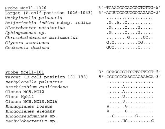
Two 16S rRNA-targeted oligonucleotide probes, Mcell-1026 and Mcell-181, were developed for specific detection of the acidophilic methanotroph Methylocella palustris using fluorescence in situ hybridization (FISH). The fluorescence signal of probe Mcell-181 was enhanced by its combined application with the oligonucleotide helper probe H158. Mcell-1026 and Mcell-181, as well as 16S rRNA oligonucleotide probes with reported group specificity for either type I methanotrophs (probes M-84 and M-705) or the Methylosinus/Methylocystis group of type II methanotrophs (probes MA-221 and M-450), were used in FISH to determine the abundance of distinct methanotroph groups in a Sphagnum peat sample of pH 4.2. M. palustris was enumerated at greater than 10(6) cells per g of peat (wet weight), while the detectable population size of type I methanotrophs was three orders of magnitude below the population level of M. palustris. The cell counts with probe MA-221 suggested that only 10(4) type II methanotrophs per g of peat (wet weight) were present, while the use of probe M-450 revealed more than 10(6) type II methanotroph cells per g of the same samples. This discrepancy was due to the fact that probe M-450 targets almost all currently known strains of Methylosinus and Methylocystis, whereas probe MA-221, originally described as group specific, does not detect a large proportion of Methylocystis strains. The total number of methanotrophic bacteria detected by FISH was 3.0 (+/-0.2) x 10(6) cells per g (wet weight) of peat. This was about 0.8% of the total bacterial cell number. Thus, our study clearly suggests that M. palustris and a defined population of Methylocystis spp. were the predominant methanotrophs detectable by FISH in an acidic Sphagnum peat bog.
Figures
References
-
- Becking J-H. Genus Beijerinckia. In: Krieg N R, Holt J G, editors. Bergey's manual of systematic bacteriology. Vol. 1. Baltimore, Md: Williams and Wilkins; 1984. pp. 311–321.
Publication types
MeSH terms
Substances
LinkOut - more resources
Full Text Sources
Molecular Biology Databases
Miscellaneous
