. 2001 Oct 1;155(1):113-22.
doi: 10.1083/jcb.200106158. Epub 2001 Sep 24.

In vivo requirement of the alpha-syntrophin PDZ domain for the sarcolemmal localization of nNOS and aquaporin-4

Affiliations

In vivo requirement of the alpha-syntrophin PDZ domain for the sarcolemmal localization of nNOS and aquaporin-4

M E Adams et al. J Cell Biol. .

Abstract

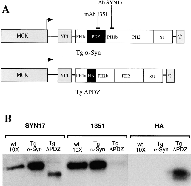
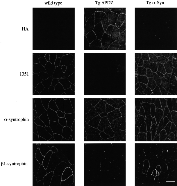
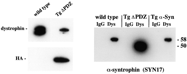
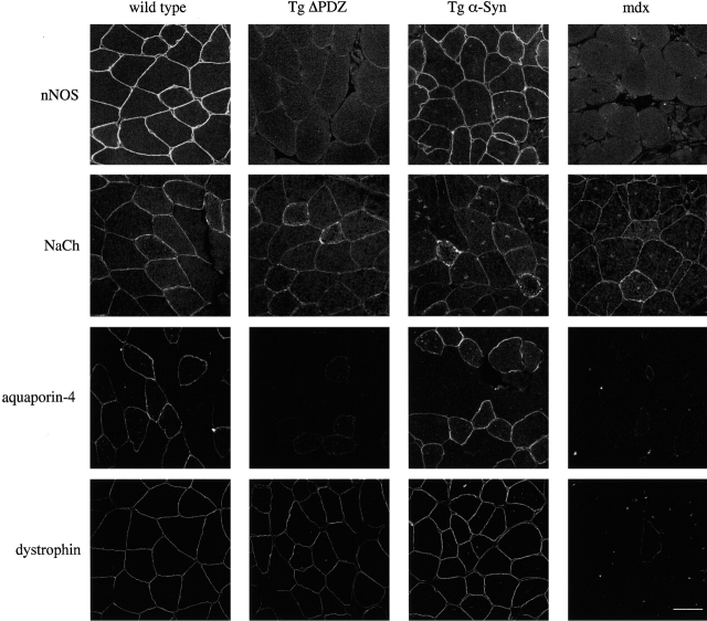
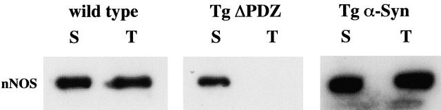
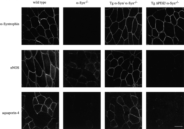
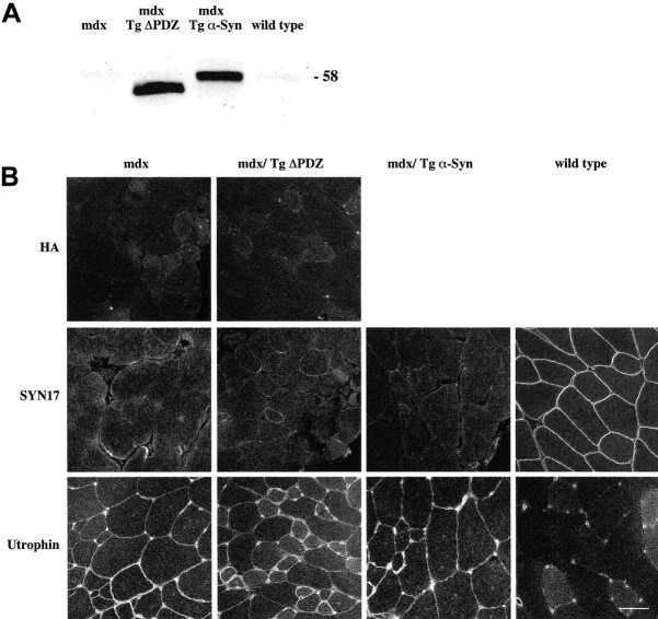
alpha-Syntrophin is a scaffolding adapter protein expressed primarily on the sarcolemma of skeletal muscle. The COOH-terminal half of alpha-syntrophin binds to dystrophin and related proteins, leaving the PSD-95, discs-large, ZO-1 (PDZ) domain free to recruit other proteins to the dystrophin complex. We investigated the function of the PDZ domain of alpha-syntrophin in vivo by generating transgenic mouse lines expressing full-length alpha-syntrophin or a mutated alpha-syntrophin lacking the PDZ domain (Delta PDZ). The Delta PDZ alpha-syntrophin displaced endogenous alpha- and beta 1-syntrophin from the sarcolemma and resulted in sarcolemma containing little or no syntrophin PDZ domain. As a consequence, neuronal nitric oxide synthase (nNOS) and aquaporin-4 were absent from the sarcolemma. However, the sarcolemmal expression and distribution of muscle sodium channels, which bind the alpha-syntrophin PDZ domain in vitro, were not altered. Both transgenic mouse lines were bred with an alpha-syntrophin-null mouse which lacks sarcolemmal nNOS and aquaporin-4. The full-length alpha-syntrophin, not the Delta PDZ form, reestablished nNOS and aquaporin-4 at the sarcolemma of these mice. Genetic crosses with the mdx mouse showed that neither transgenic syntrophin could associate with the sarcolemma in the absence of dystrophin. Together, these data show that the sarcolemmal localization of nNOS and aquaporin-4 in vivo depends on the presence of a dystrophin-bound alpha-syntrophin PDZ domain.


Figures

Figure 1.
Figure 1.
Construction and characterization of transgenic mice. (A) The mouse cDNA sequence of full-length α-syntrophin and α-syntrophin missing the PDZ domain (ΔPDZ) was placed downstream of the mouse muscle creatine kinase (MCK) promoter and the VP1 intron of plasmid pCKVA. The PDZ domain of the Tg ΔPDZ construct was replaced with an HA tag. The binding sites for mAb 1351 and Ab SYN 17 are shown. (B) Expression of transgene products was examined by immunoblotting. 20 μg (wild-type control) and 2 μg (transgenics) of total protein isolated from skeletal muscle were subjected to immunoblot analysis. SYN17 recognizes both endogenous and transgenic α-syntrophin; mAb 1351 recognizes endogenous and Tg α-Syn but not Tg ΔPDZ; HA recognizes only Tg ΔPDZ. Very small amounts of endogenous α-syntrophin can be seen in the Tg ΔPDZ preparations with SYN17 and mAb 1351.
Figure 2.
Figure 2.
Comparative localization of α-syntrophin in control and transgenic mice. Immunofluorescence microscopy of adult (>8 wk) mouse quadriceps muscle shows that the HA-tagged Tg ΔPDZ transgene product is localized at the sarcolemma. Labeling with mAb 1351 is strong on normal and Tg α-Syn muscle but shows greatly reduced intensity in the Tg ΔPDZ muscle. Thus, Tg ΔPDZ syntrophin displaces the endogenous α-syntrophin. Antibody SYN17 that recognizes both transgene products and endogenous α-syntrophin shows a modest increase in labeling intensity at the sarcolemma and in the cytosol in the transgenic muscle. β1-Syntrophin is displaced by the high expression of the Tg ΔPDZ product but remains present, although at reduced levels, in the muscle of the full-length α-syntrophin transgenic mouse. β1-Syntrophin is also detected in blood vessels surrounding muscle fibers in all sections. Bar, 50 μm.
Figure 3.
Figure 3.
Association of the Tg ΔPDZ product with dystrophin. Dystrophin and associated proteins were immunoprecipitated from wild-type (C57) and Tg ΔPDZ skeletal muscle (Dys). Control samples were prepared by substituting normal mouse IgG for the dystrophin antibody (IgG). (A) Immunoblotting with the HA antibody shows that the transgene product copurifies with dystrophin. (B) Immunoblotting with SYN17 confirms that the Tg ΔPDZ product displaces endogenous syntrophin from the dystrophin complex (no 58-kD band, corresponding to the full-length endogenous α-syntrophin, is seen in the Tg ΔPDZ lane).
Figure 4.
Figure 4.
Immunofluorescence analysis of syntrophin-associated proteins in mouse quadriceps muscle. In the absence of the α-syntrophin PDZ domain (Tg ΔPDZ) or dystrophin (mdx), nNOS and aquaporin-4 fail to localize at the sarcolemma while sodium channel (NaCh) distribution appears unchanged. Dystrophin levels appear normal in the transgenic animals. Note: the dystrophin-labeled fiber in the mdx mouse is probably a revertant fiber and serves as a convenient focussing and antibody control. Bar, 50 μm.
Figure 5.
Figure 5.
nNOS is shifted from cytoskeletal association to the soluble pool in the Tg ΔPDZ muscle. Samples from soluble cytosolic (S) or 1% Triton (T) skeletal muscle extracts were analyzed for nNOS by immunoblotting. In the absence of the α-syntrophin PDZ domain, nNOS is found only in the soluble fraction. High levels of expression of full-length α-syntrophin do not change the relative amounts of nNOS in the cytosolic and insoluble fractions.
Figure 6.
Figure 6.
Sarcolemmal expression of nNOS and aquaporin-4 in genetic crosses of α-syntrophin–null mice with transgenic mice. Both nNOS and aquaporin-4 are absent from the sarcolemma of α-syntrophin–null mice. Muscle from α-syntrophin–null mice crossed with Tg α-Syn mice shows sarcolemmal expression of both nNOS and aquaporin-4. Sarcolemmal expression of these proteins is not restored in muscle of α-syntrophin–null mice crossed with Tg ΔPDZ mice. Bar, 50 μm.
Figure 7.
Figure 7.
Distribution of Tg ΔPDZ and Tg α-Syn in the absence of dystrophin. (A) Both Tg ΔPDZ and Tg α-Syn proteins (detected with SYN17) are expressed at high levels in muscle lacking dystrophin (mdx/Tg ΔPDZ and mdx/Tg α-Syn). (B) Despite high levels of transgene product expression, neither the full-length nor the Tg ΔPDZ syntrophin are found on the sarcolemma. Utrophin sarcolemmal expression is upregulated in the mdx mouse but apparently to levels insufficient to recruit α-syntrophin. Bar, 50 μm.

References

    1. Abdelmoity, A., R.C. Padre, K.E. Burzynski, J.T. Stull, and K.S. Lau. 2000. Neuronal nitric oxide synthase localizes through multiple structural motifs to the sarcolemma in mouse myotubes. FEBS Lett. 482:65–70. - PubMed
    1. Adams, M.E., M.H. Butler, T.M. Dwyer, M.F. Peters, A.A. Murnane, and S.C. Froehner. 1993. Two forms of mouse syntrophin, a 58 kd dystrophin-associated protein, differ in primary structure and tissue distribution. Neuron. 11:531–540. - PubMed
    1. Adams, M.E., T.M. Dwyer, L.L. Dowler, R.A. White, and S.C. Froehner. 1995. Mouse alpha 1- and beta 2-syntrophin gene structure, chromosome localization, and homology with a discs large domain. J. Biol. Chem. 270:25859–25865. - PubMed
    1. Adams, M.E., N. Kramarcy, S.P. Krall, S.G. Rossi, R.L. Rotundo, R. Sealock, and S.C. Froehner. 2000. Absence of alpha-syntrophin leads to structurally aberrant neuromuscular synapses deficient in utrophin. J. Cell Biol. 150:1385–1398. - PMC - PubMed
    1. Ahn, A.H., and L.M. Kunkel. 1995. Syntrophin binds to an alternatively spliced exon of dystrophin. J. Cell Biol. 128:363–371. - PMC - PubMed

Publication types

MeSH terms