. 2001 Sep 6;20(39):5431-9.
doi: 10.1038/sj.onc.1204719.

HPV E6 and MAGUK protein interactions: determination of the molecular basis for specific protein recognition and degradation

Affiliations

HPV E6 and MAGUK protein interactions: determination of the molecular basis for specific protein recognition and degradation

M Thomas et al. Oncogene. .

Abstract

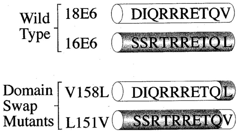
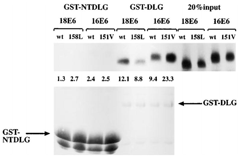
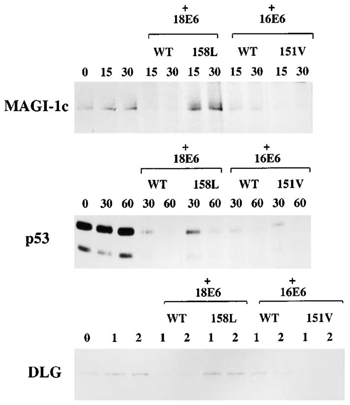
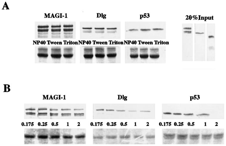
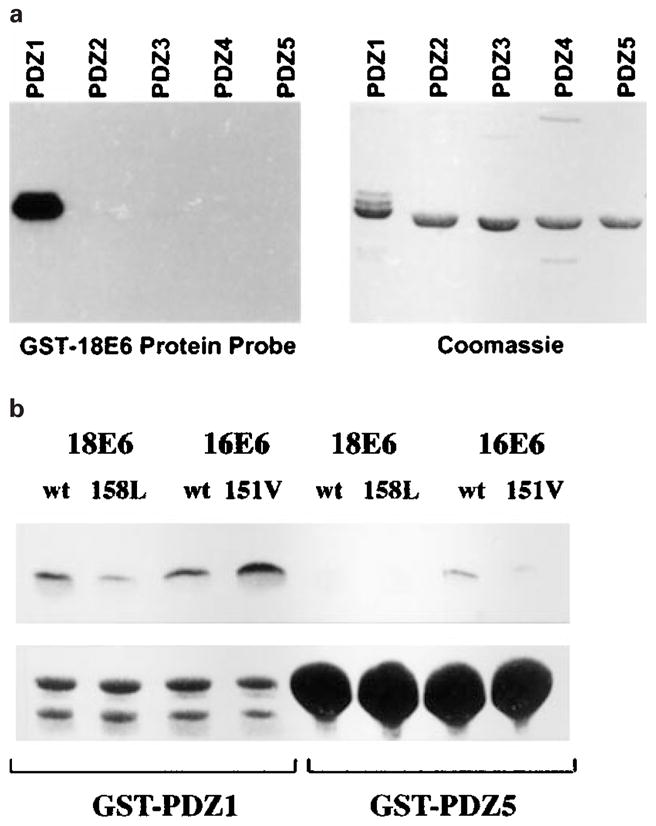
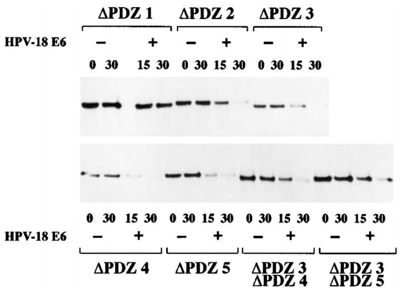
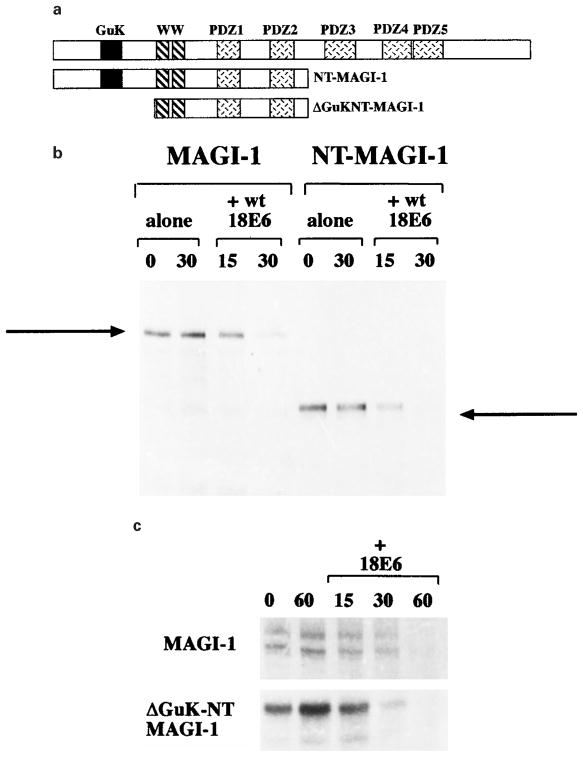
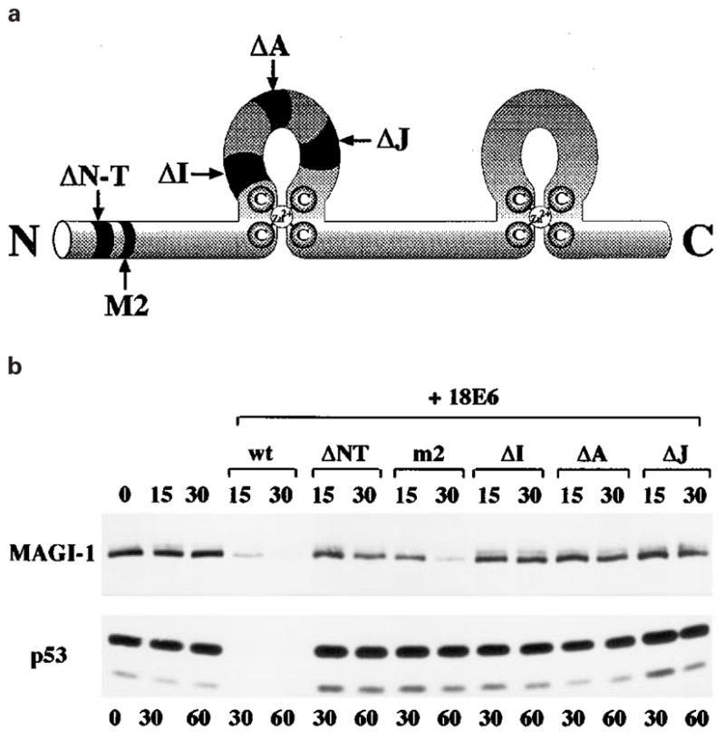
It has recently been shown that the high-risk human papillomavirus (HPV) E6 proteins can target the PDZ-domain containing proteins, Dlg, MUPP-1, MAGI-1 and hScrib for proteasome-mediated degradation. However, the E6 proteins from HPV-16 and HPV-18 (the two most common high-risk virus types) differ in their ability to target these proteins in a manner that correlates with their malignant potential. To investigate the underlying mechanisms for this, we have mutated HPV-16 and HPV-18 E6s to give each protein the other's PDZ-binding motif. Analysis of these mutants shows that the greater ability of HPV-18 E6 to bind to these proteins and to target them for degradation is indeed due to a single amino acid difference. Using a number of assays, we show that the E6 proteins interact specifically with only one of the five PDZ domains of MAGI-1, and this is the first interaction described for this particular PDZ domain. We also show that the guanylate kinase homology domain and the regions of MAGI-1 downstream of amino acid 733 are not required for the degradation of MAGI-1. Finally, in a series of comparative analyses, we show that the degradation of MAGI-1 occurs through a different mechanism from that used by the E6 protein to induce the degradation of Dlg and p53.


Figures

Figure 1
Figure 1
Alignment of the carboxy-terminal amino acid residues of wild type and mutant HPV-16 and HPV-18 E6 proteins
Figure 2
Figure 2
The domain-swap mutants of E6 differ in their ability to bind GST-DLG in vitro. The upper panel shows 20% of input in vitro translated E6 proteins, together with that retained on either GST-NTDLG or GST-DLG. The numbers indicate the percentage of load retained. The lower panel shows the same gel rehydrated and stained with Coomassie Blue to control GST fusion protein input
Figure 3
Figure 3
In vitro degradation of E6 target proteins with the domain swap mutants. In vitro translated wild type and mutant E6 proteins were incubated with in vitro translated MAGI-1, DLG or p53. The MAGI-1 assay was incubated for 15 and 30 min, the p53 assay for 30 and 60 min, and the DLG assay for 1 and 2 h. In each case, the remaining target protein was detected by immunoprecipitation, followed by SDS–PAGE and auto-radiography
Figure 4
Figure 4
Analysis of the binding affinities between GST-HPV-18 E6 and the E6 target proteins. (a) Binding affinities in different detergents. The upper panels show GST pull-down assays using GST-18E6 and in vitro translated MAGI-1, Dlg and p53. The samples were each washed with 2% of the detergent shown, in PBS. Twenty per cent of the respective input protein are shown in the right hand panel. The lower panels show the rehydrated and Coomassie-stained gels. (b) Binding affinities at increasing concentrations of NaCl. Similar binding assays were washed with either PBS alone (0.175 M NaCl), or with increasing concentrations (M) of NaCl, as indicated. The lower panels show the rehydrated and Coomassie-stained gels
Figure 5
Figure 5
GST-18E6 binds specifically to GST-PDZ domain 1 of MAGI-1. (a) GST fusion proteins were constructed, consisting of each individual PDZ domain of MAGI-1. They were purified, separated by SDS –PAGE and blotted onto nitrocellulose membrane, which was then probed with a purified, radiolabelled GST-HPV18 E6 fusion protein. Bound radiolabelled protein was detected by autoradiography. (b) A GST pull-down assay was performed using GST-PDZ1 and GST-PDZ5 proteins, together with in vitro translated wild type HPV-18 and HPV-16 E6s, and the domain-swap mutants E6. The percentage of wt 18E6, V158L, wt 16E6, L151V loads retained were 14.3, 6.7, 10.8 and 22.1% respectively on GST-PDZ1, and 2.5, 2.4, 3.2 and 2.6% respectively on GST-PDZ5
Figure 6
Figure 6
In vitro degradation of MAGI-1 requires PDZ domain 1. The MAGI-1b PDZ deletion mutants were translated in vitro, and were then incubated at 30°C for the time/minutes indicated, with or without HPV-18 E6. The remaining protein was detected by immunoprecipitation and autoradiography, as before
Figure 7
Figure 7
Only the amino terminal half of MAGI-1 is required for HPV-18 E6-induced degradation. (a) A schematic diagram of the MAGI-1 deletion mutants. (b) Wild type MAGI-1 and the NT-MAGI-1 mutant were translated in vitro and in vitro degradation assays were performed in the presence of HPV-18 E6 for 15 and 30 min. (c) Wild type MAGI-1 and the ΔGuK-NT-MAGI-1 mutant were translated in vitro and in vitro degradation assays were performed as in b
Figure 8
Figure 8
HPV-18 E6 induces the degradation of MAGI-1 by a different mechanism from that used in the degradation of p53. (a) A schematic diagram of the HPV-18 E6 protein, showing the positions of the deletion mutants used. (b) A comparison of the in vitro degradation of p53 and of MAGI-1 induced by the deletion mutants of HPV-18 E6. MAGI-1 was incubated with E6 for 15 and 30 min, while p53 was incubated for 30 and 60 min

References

    1. Banks L, Matlashewski G, Crawford L. Eur J Biochem. 1986;159:529–534. - PubMed
    1. Bilder D, Li M, Perrimon N. Science. 2000;289:113–116. - PubMed
    1. Craven S, Bredt D. Cell. 1998;93:495–498. - PubMed
    1. Crook T, Tidy J, Vousden K. Cell. 1991a;67:547–556. - PubMed
    1. Crook T, Wrede D, Vousden KH. Oncogene. 1991b;6:873–875. - PubMed

Publication types

MeSH terms