. 2001 Oct 9;98(21):12150-5.
doi: 10.1073/pnas.201407398. Epub 2001 Sep 25.

Soluble HLA-G protein secreted by allo-specific CD4+ T cells suppresses the allo-proliferative response: a CD4+ T cell regulatory mechanism

Affiliations

Soluble HLA-G protein secreted by allo-specific CD4+ T cells suppresses the allo-proliferative response: a CD4+ T cell regulatory mechanism

N Lila et al. Proc Natl Acad Sci U S A. .

Abstract

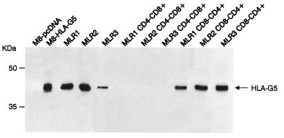
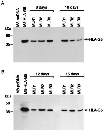
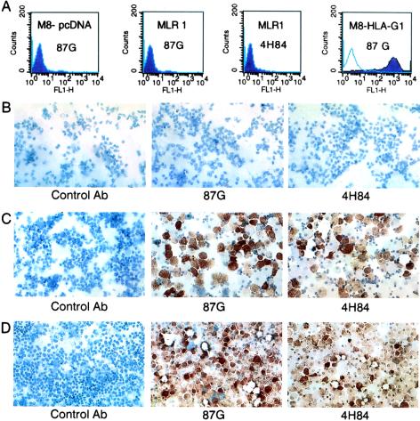
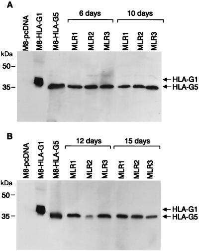
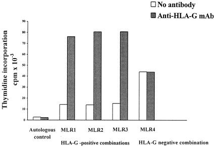
We recently reported that the nonclassical HLA class I molecule HLA-G was expressed in the endomyocardial biopsies and sera of 16% of heart transplant patients studied. The aim of the present report is to identify cells that may be responsible for HLA-G protein expression during the allogeneic reaction. Carrying out mixed lymphocyte cultures in which the responder cell population was depleted either in CD4(+) or CD8(+) T cells, we found that soluble HLA-G5 protein but not the membrane-bound HLA-G isoform was secreted by allo-specific CD4(+) T cells from the responder population, which suppressed the allogeneic proliferative T cell response. This inhibition may be reversed by adding the anti-HLA-G 87G antibody to a mixed lymphocyte culture. That may indicate a previously uncharacterized regulatory mechanism of CD4(+) T cell proliferative response.


Figures

Figure 1
Figure 1
Immunoprecipitation followed by Western blot analysis of cell culture supernatants after 10 days of MLR revealed the presence of soluble HLA-G5 in three MLR combinations (MLR1, MLR2, and MLR3). Immunoprecipitation and Western blot analysis of soluble HLA-G were carried out by using the PAG5-6 Ab and 4H84 mAb, respectively. In these HLA-G5-positive combinations, when the responder cells were CD4+-depleted (MLR1 CD4CD8+, MLR2 CD4CD8+, and MLR3 CD4CD8+), HLA-G5 could not be detected. In contrast, when responder cells were CD8+-depleted (MLR1 CD8CD4+, MLR2 CD8CD4+, and MLR3 CD8CD4+), HLA-G5 could be observed. M8-HLA-G5 and M8-pcDNA transfectant cells were used as positive and negative controls, respectively.
Figure 2
Figure 2
Detection of HLA-G5 by immunoprecipitation followed by Western blot analysis of cell culture supernatants on various days after MLR. Stimulator cells were used against total peripheral blood mononuclear responder cells in the three HLA-G-positive combinations (MLR1, MLR2, and MLR3). (A) Days 6 and 10 of primary MLR. (B) Days 12 and 15 of secondary MLR. M8-HLA-G5 transfectant cells were used as the positive control, and M8-pcDNA was used as the negative control.
Figure 3
Figure 3
Flow cytometry and immunocytochemical analysis after 10 days of MLR by using the 87G and 4H84 anti-HLA-G mAbs. (A) Flow cytometry profiles were all negative with anti-HLA-G mAbs, whereas HLA-G1-transfected cells were positively stained with 87G. (B) Immunocytochemical analysis was carried out without permeabilization. In this HLA-G-positive combination, the PBMCs were negatively stained; after permeabilization, the responder cells correspond to all of the PBMCs (C) and CD8+-depleted PBMCs (D) were positively stained.
Figure 4
Figure 4
Western blot analysis of responder cell lysates after MLR. Stimulator cells were used against total peripheral blood mononuclear responder cells in the three HLA-G-positive combinations (MLR1, MLR2, and MLR3). (A) Days 6 and 10 of primary MLR. (B) Days 12 and 15 of secondary MLR. M8-HLA-G5 and M8-HLA-G1 transfectant cells were used as positive controls, and M8-pcDNA was used as the negative control.
Figure 5
Figure 5
Effect of HLA-G5 production on T cell proliferation when (i) the responder cells were CD8+-depleted (CD4+-enriched) and (ii) the anti-HLA-G5 mAb was used in HLA-G-positive and -negative combinations. The data are expressed as the mean of thymidine incorporation (cpm) in triplicate wells.

References

    1. Kovats S, Main E K, Librach C, Stubblebine M, Fisher S J, De Mars R. Science. 1990;248:220–223. - PubMed
    1. McMaster M T, Librach C, Zhou Y, Lim K H, Janatpour M J, De Mars R, Kovats S, Damsky C, Fisher S J. J Immunol. 1995;154:3771–3778. - PubMed
    1. Rouas-Freiss N, Marchal-Bras Goncalves R, Menier C, Dausset J, Carosella E D. Proc Natl Acad Sci USA. 1997;94:11520–11525. - PMC - PubMed
    1. Ishitani A, Geraghty D E. Proc Natl Acad Sci USA. 1992;89:3947–3951. - PMC - PubMed
    1. Fujii T, Ishitani A, Geraghty D E. J Immunol. 1994;153:5516–5524. - PubMed

Publication types

MeSH terms

LinkOut - more resources