. 2001 Oct 9;98(21):12221-6.
doi: 10.1073/pnas.211106398. Epub 2001 Sep 25.

Microinjection and growth of bacteria in the cytosol of mammalian host cells

Affiliations

Microinjection and growth of bacteria in the cytosol of mammalian host cells

M Goetz et al. Proc Natl Acad Sci U S A. .

Abstract

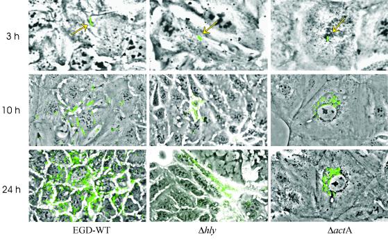
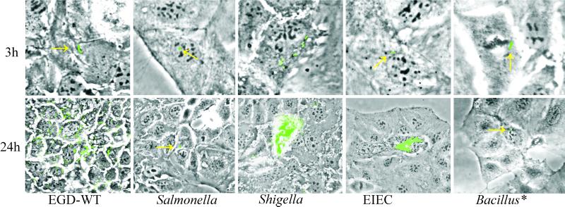
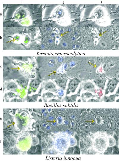
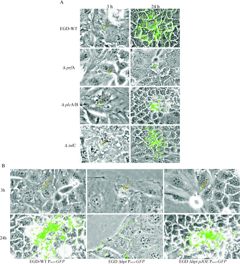
Most facultative intracellular bacteria replicate in specialized phagosomes after being taken up by mammalian cells. Relatively few intracellular bacteria escape the phagosomal compartment with the help of cytolytic (pore-forming) proteins and replicate in the host cell cytosol. Without such toxins, intracellular bacteria cannot reach this cellular compartment. To circumvent the requirement of an "escape" step, we developed a procedure allowing the efficient direct injection of bacteria into the cytosol of mammalian cells. With this technique, we show that most bacteria, including extracellular bacteria and intracellular pathogens that normally reside in a vacuole, are unable to replicate in the cytosol of the mammalian cells. In contrast, microorganisms that replicate in the cytosol, such as Listeria monocytogenes, Shigella flexneri, and, to some extent, enteroinvasive Escherichia coli, are able to multiply in this cellular compartment after microinjection. Further L. monocytogenes with deletion in its PrfA-regulated hpt gene was found to be impaired in replication when injected into the cytosol. Complementation of the hpt mutation with a plasmid carrying the wild-type hpt gene restored the replication ability in the cytosol. These data indicate that cytosolic intracellular pathogens have evolved specific mechanisms to grow in this compartment of mammalian cells.


Figures

Figure 1
Figure 1
Microinjection of wild-type (WT) and mutant strains (Δhly and ΔactA) of L. monocytogenes into Caco-2 cells. Microinjection into Caco-2 cells was performed as described in Materials and Methods, using L. monocytogenes wild-type strain EGD and isogenic mutants with deletions in hly and actA. All three strains harbor the same plasmid carrying PactA-gfp. Bacterial replication was determined after 3, 10, and 24 h. Arrows mark single fluorescent bacteria, which appear 3 h after microinjection.
Figure 2
Figure 2
Microinjection of Caco-2 cells with L. monocytogenes EGD, S. typhimurium 14028s, S. flexneri iscA, EIEC strain W7062, and B. subtilis DSM401. All strains were GFP-labeled by transformation with pKSBC16-Psod-gfp. Bacteria grown overnight were used for microinjection. Intracellular bacterial replication was monitored until 24 h after injection. Photographs shown were taken at 3 and 24 h postinjection, except for B. subtilis (Bacillus*), which were taken at 1 and 6 h postinjection; in this case, there were hardly any fluorescent bacteria after 24 h.
Figure 3
Figure 3
Atypical replication of Y. enterocolitica, B. subtilis, and L. innocua, each carrying the Psod-gfp plasmid in Caco-2 cells after microinjection. With a frequency of about 1 in 30 successfully microinjected cells, extensive replication of the bacteria were observed 12 to 24 h postinjection. These host cells were apoptotic or necrotic as demonstrated by staining with Hoechst 33342 (20 μg/ml) and propidium iodide (2 μg/ml) in contrast to those cells that carried a single bacterium. Note the lower magnification of panels 2b, 2c, 2e and 3b, 3c, 3e, which was used to visualize neighboring cells with intact nuclei for comparison. The Caco-2 cell shown in panel 1d has already lysed to some extent, and the fluorescent bacteria seen at the neighboring cells are not intracellular. This is occasionally seen when the host cells become necrotic. Arrows point to the same cells before and after staining with the Hoechst 33342 (panels 2a–2f) and propidium iodide (panels 3a–3f).
Figure 4
Figure 4
(A) Microinjection of Caco-2 cells with L. monocytogenes EGD and the isogenic mutants with in-frame deletions in the PrfA-regulated genes, prfA, plcA, and plcB, and inlC and (B) with a mutant carrying an in-frame deletion in hpt gene and with this mutant complemented with a plasmid carrying the hpt gene under the control of its own PrfA-dependent promoter together with the PactA-regulated gfp cassette (pJOE-PactA-gfp). The isogenic EGD wild-type strain carrying a similar plasmid without the hpt gene was used as control. Intracellular bacterial replication was monitored for 24 h; photographs of microinjected Caco-2 cells shown were taken after 3 h and 24 h.

References

    1. Cossart P, Lecuit M. EMBO J. 1998;17:3797–3806. - PMC - PubMed
    1. Kuhn M, Goebel W. Immunol Rev. 1997;158:57–67. - PubMed
    1. Finlay B, Falkow S. Microbiol Mol Biol Rev. 1997;61:136–169. - PMC - PubMed
    1. Galan J E. Mol Microbiol. 1996;20:263–271. - PubMed
    1. Camilli A, Goldfine H, Portnoy D A. J Exp Med. 1991;173:751–754. - PMC - PubMed

Publication types

MeSH terms

Substances

LinkOut - more resources