. 2001 Oct 9;98(21):12301-6.
doi: 10.1073/pnas.211209098. Epub 2001 Sep 25.

"What" and "where" in the human auditory system

Affiliations

"What" and "where" in the human auditory system

C Alain et al. Proc Natl Acad Sci U S A. .

Abstract

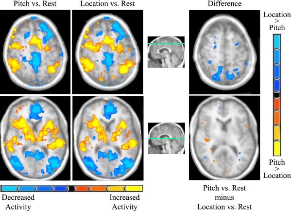
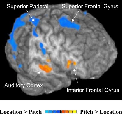
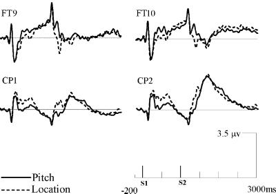
The extent to which sound identification and sound localization depend on specialized auditory pathways was examined by using functional magnetic resonance imaging and event-related brain potentials. Participants performed an S1-S2 match-to-sample task in which S1 differed from S2 in its pitch and/or location. In the pitch task, participants indicated whether S2 was lower, identical, or higher in pitch than S1. In the location task, participants were asked to localize S2 relative to S1 (i.e., leftward, same, or rightward). Relative to location, pitch processing generated greater activation in auditory cortex and the inferior frontal gyrus. Conversely, identifying the location of S2 relative to S1 generated greater activation in posterior temporal cortex, parietal cortex, and the superior frontal sulcus. Differential task-related effects on event-related brain potentials (ERPs) were seen in anterior and posterior brain regions beginning at 300 ms poststimulus and lasting for several hundred milliseconds. The converging evidence from two independent measurements of dissociable brain activity during identification and localization of identical stimuli provides strong support for specialized auditory streams in the human brain. These findings are analogous to the "what" and "where" segregation of visual information processing, and suggest that a similar functional organization exists for processing information from the auditory modality.


Figures

Figure 1
Figure 1
The first two columns (Left) show the group mean activation during the pitch and location tasks vs. rest, respectively. The color scale below is based on t values ranging from 6 to 15 (P < 0.00001). The rightmost column shows the difference in brain activation between the location and the pitch tasks. The color scale for this image is based on t values ranging from 2.9 to 7 (P < 0.01). Areas with greater activity during the location task are shown in blue, whereas those more active during the pitch task are illustrated in orange and yellow. The right hemisphere is shown on the left side of the images. Slice locations are indicated by green lines on the accompanying midline sagittal image.
Figure 2
Figure 2
Three-dimensional pattern of cortical activation to highlight differences between pitch and location discrimination tasks. The right hemisphere is foremost in the figure and part of the temporal lobe has been removed to show activation in temporal and inferior frontal cortices. The enhanced signal in posterior visual areas during the location task was caused by greater signal reduction during the pitch task rather than increased activity during the location task. See Table 1 for atlas coordinates and statistical measures for these areas. The color scale for this image is based on t values ranging from 2.9 to 7 (P < 0.01).
Figure 3
Figure 3
Group mean event-related waveforms elicited during the pitch (solid line) and the location (dashed line) judgment tasks. FT9/FT10, left and right frontal-temporal electrodes; CP1/CP2, left and right central-parietal electrodes.

References

    1. Rauschecker J P, Tian B, Hauser M. Science. 1995;268:111–114. - PubMed
    1. Rauschecker J P, Tian B, Pons T, Mishkin M. J Comp Neurol. 1997;382:89–103. - PubMed
    1. Rauschecker J P. Audiol Neurootol. 1998;3:86–103. - PubMed
    1. Hackett T A, Stepniewska I, Kaas J H. Brain Res. 1999;817:45–58. - PubMed
    1. Romanski L M, Tian B, Fritz J, Mishkin M, Goldman-Rakic P S, Rauschecker J P. Nat Neurosci. 1999;2:1131–1136. - PMC - PubMed

Publication types

LinkOut - more resources