. 2001 Oct 9;98(21):11955-60.
doi: 10.1073/pnas.211305198. Epub 2001 Sep 25.

An expanded glutamine repeat destabilizes native ataxin-3 structure and mediates formation of parallel beta -fibrils

Affiliations

An expanded glutamine repeat destabilizes native ataxin-3 structure and mediates formation of parallel beta -fibrils

A E Bevivino et al. Proc Natl Acad Sci U S A. .

Abstract

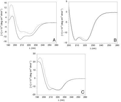
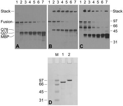
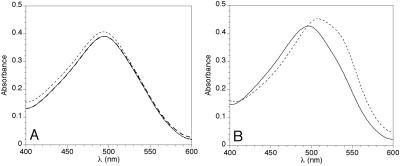
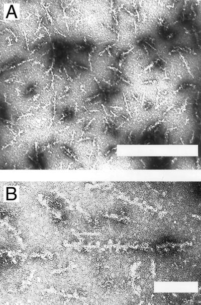
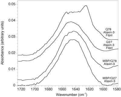
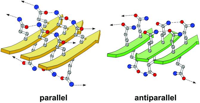
The protein ataxin-3 contains a polyglutamine region; increasing the number of glutamines beyond 55 in this region gives rise to the neurodegenerative disease spinocerebellar ataxia type 3. This disease and other polyglutamine expansion diseases are characterized by large intranuclear protein aggregates (nuclear inclusions). By using full-length human ataxin-3, we have investigated the changes in secondary structure, aggregation behavior, and fibril formation associated with an increase from the normal length of 27 glutamines (Q27 ataxin-3) to a pathogenic length of 78 glutamines (Q78 ataxin-3). Q78 ataxin-3 aggregates strongly and could be purified only when expressed with a solubility-enhancing fusion-protein partner. A marked decrease in alpha-helical secondary structure accompanies expansion of the polyglutamine tract, suggesting destabilization of the native protein. Proteolytic removal of the fusion partner in the Q78 protein, but not in the Q27 protein, leads to the formation of SDS-resistant aggregates and Congo-red reactive fibrils. Infrared spectroscopy of fibrils reveals a high beta-sheet content and suggests a parallel, rather than an antiparallel, sheet conformation. We present a model for a polar zipper composed of parallel polyglutamine beta-sheets. Our data show that intact ataxin-3 is fully competent to form aggregates, and posttranslational cleavage or other processing is not necessary to generate a misfolding event. The data also suggest that the protein aggregation phenotype associated with glutamine expansion may derive from two effects: destabilization of the native protein structure and an inherent propensity for beta-fibril formation on the part of glutamine homopolymers.


Figures

Figure 1
Figure 1
CD spectra of various ataxin-3 species. (A) Solid line, hexahistidine-Q27 ataxin-3; dotted line, MBP. (B) Solid line, MBP/Q27 ataxin-3 fusion protein; dotted line, calculated spectrum obtained from a mass-weighted average of the MBP and Q27 ataxin-3 spectra found in A. (C) Solid line, MBP/Q27 ataxin-3 fusion; dotted line, MBP/Q78 ataxin-3 fusion.
Figure 2
Figure 2
(AC) Western blots showing the time course of TEV protease digestion of MBP/ataxin-3 fusion proteins. (A) Digestion of MBP/Q27 ataxin-3. (B and C) Digestion of MBP/Q78 ataxin-3. A and B are probed with polyclonal anti-ataxin-3; C is probed with anti-MBP. Lane 1: time = 0; lane 2: 15 min; lane 3: 30 min; lane 4: 1 hr; lane 5: 2 hr; lane 6: 3 hr; lane 7: 4 hr. The high-molecular-weight species seen in B and C have failed to enter the resolving gel and remain in the stacking gel. (D) Coomassie-stained SDS/PAGE gel of MBP/ataxin-3 fusion proteins. Lane 1: MBP/Q27 ataxin-3; lane 2: MBP/Q78 ataxin-3; lane M: molecular-weight markers.
Figure 3
Figure 3
CR binding by MBP/ataxin-3 fusion proteins, before and after exposure to fibril-growth conditions. TEV protease was added at day 0, and fusion proteins were incubated at 37°C, as described under Materials and Methods. (A) MBP/Q27 ataxin-3 fusion. CR only (no protein added), thick dashed line; solid line, day 0; dotted line, day 5. No significant dye-binding by the protein is observed at either day 0 or day 5. In fact, the figure shows that the spectra for CR alone and for protein at day 0 are virtually identical. (B) MBP/Q78 ataxin-3 fusion. Solid line, day 0; dotted line, day 5. A marked increase in dye-binding is seen to accompany fibril formation at day 5.
Figure 4
Figure 4
Electron micrographs of negatively stained Q78/ataxin-3 fibrils at (A) 33,000× (Bar = 500 nm) and (B) 100,000× magnification (Bar = 100 nm).
Figure 5
Figure 5
Infrared spectra of intact MBP/ataxin-3 fusion proteins and of cleaved fusion proteins exposed to fibril-forming conditions. Transmission spectra are shown for intact fusions, and internal reflection spectra are shown for cleaved and fibrillized fusions.
Figure 6
Figure 6
Molecular models of homopolymeric glutamine peptides forming either parallel β-sheets (gold) or antiparallel β-sheets (green). Models containing four 11-residue strands were built in QUANTA (Accelrys, San Diego) and energy-minimized with CHARMM (Harvard Univ., Cambridge, MA; ref. 37); only portions of the models are shown for the sake of clarity. The parallel model was constructed starting from a canonical parallel β-sheet architecture followed by a search for optimal side-chain torsion angles. The antiparallel model is based upon figure 1 in ref. . The strands of the β-sheet are represented by solid arrows, and the glutamine side chains are shown in a ball-and-stick rendering, with carbon atoms colored in gray, nitrogen atoms in blue, and oxygen atoms in red. Dashed lines represent hydrogen bonds between side chains. Arrows represent hydrogen bonds to adjacent strands that are not included in the figure. The figure was made with the program MOLSCRIPT (38).

References

    1. Zoghbi H Y, Orr H T. Annu Rev Neurosci. 2000;23:217–247. - PubMed
    1. Cummings C J, Zoghbi H Y. Hum Mol Genet. 2000;9:909–916. - PubMed
    1. Paulson H L, Das S S, Crino P B, Perez M K, Patel S C, Gotsdiner D, Fischbeck K H, Pittman R N. Ann Neurol. 1997;41:453–462. - PubMed
    1. Paulson H L. Brain Pathol. 2000;10:293–299. - PMC - PubMed
    1. Zoghbi H Y, Orr H T. Curr Opin Neurobiol. 1999;9:566–570. - PubMed

Publication types

LinkOut - more resources