Review
. 2001 Sep 25;98(20):11003-8.
doi: 10.1073/pnas.191351498.

Presynaptic kainate receptors at hippocampal mossy fiber synapses

Affiliations
Review

Presynaptic kainate receptors at hippocampal mossy fiber synapses

D Schmitz et al. Proc Natl Acad Sci U S A. .

Abstract

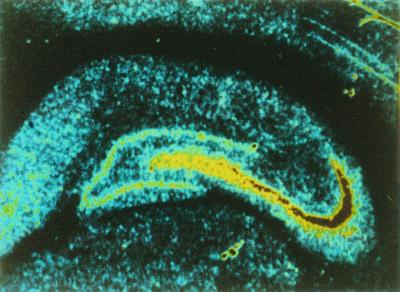
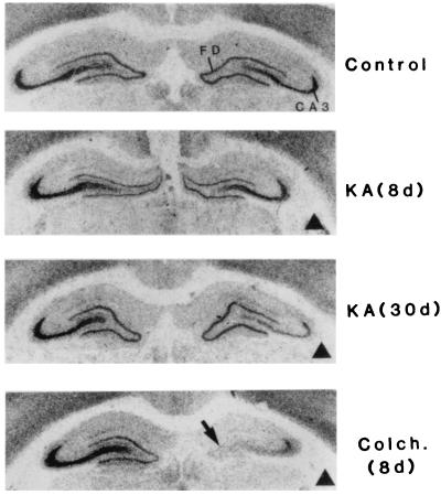
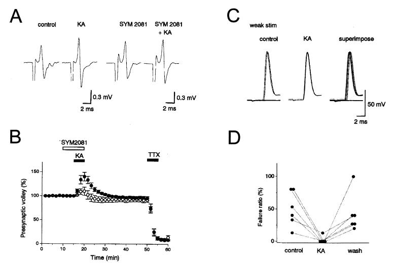
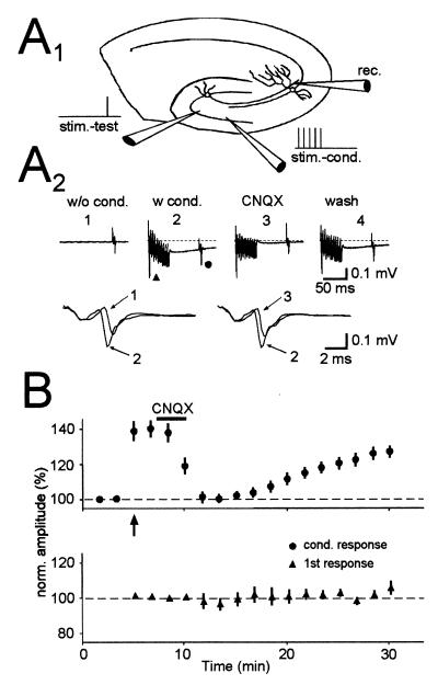
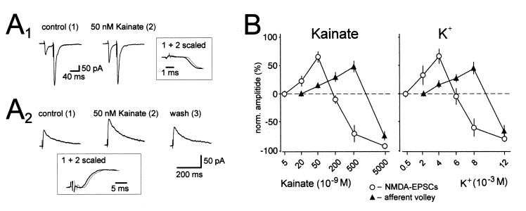
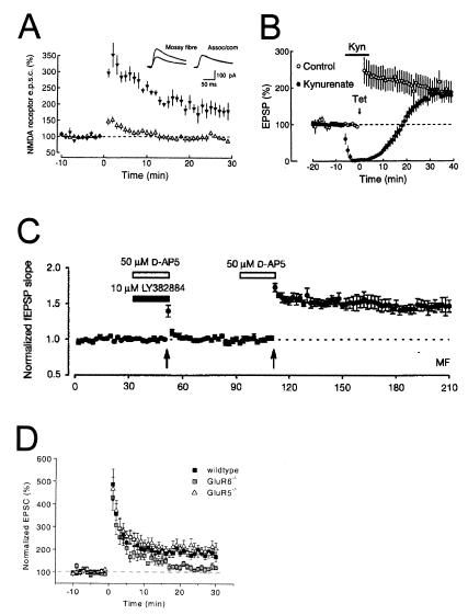
Hippocampal mossy fibers, which are the axons of dentate granule cells, form powerful excitatory synapses onto the proximal dendrites of CA3 pyramidal cells. It has long been known that high-affinity binding sites for kainate, a glutamate receptor agonist, are present on mossy fibers. Here we summarize recent experiments on the role of these presynaptic kainate receptors (KARs). Application of kainate has a direct effect on the amplitude of the extracellularly recorded fiber volley, with an enhancement by low concentrations and a depression by high concentrations. These effects are mediated by KARs, because they persist in the presence of the alpha-amino-3-hydroxy-5-methyl-4-isoxazolepropionic acid receptor-selective antagonist GYKI 53655, but are blocked by the alpha-amino-3-hydroxy-5-methyl-4-isoxazolepropionic acid/KAR antagonist 6-cyano-7-nitroquinoxaline-2,3-dione and the KAR antagonist SYM2081. The effects on the fiber volley are most likely caused by a depolarization of the fibers via the known ionotropic actions of KARs, because application of potassium mimics the effects. In addition to these effects on fiber excitability, low concentrations of kainate enhance transmitter release, whereas high concentrations depress transmitter release. Importantly, the synaptic release of glutamate from mossy fibers also activates these presynaptic KARs, causing an enhancement of the fiber volley and a facilitation of release that lasts for many seconds. This positive feedback contributes to the dramatic frequency facilitation that is characteristic of mossy fiber synapses. It will be interesting to determine how widespread facilitatory presynaptic KARs are at other synapses in the central nervous system.


Figures

Figure 1
Figure 1
Distribution of kainate binding sites in the hippocampus. Binding site density is color-coded with high to low densities represented by red-yellow-blue. The autoradiography was carried out with [3H]kainate and shows a high labeling density localized to the stratum lucidum, the termination zone for mossy fibers. [Reprinted with permission from ref. (Copyright 1982, Elsevier Science).]
Figure 2
Figure 2
Effects of selective neuronal lesions on the high-affinity kainate binding in the hippocampus. Shown are the distribution of kainate binding in (perfused) controls, kainate (KA)- or colchcine (Colch.)-treated cases. Dark triangles indicate the side ipsilateral to injection. In perfused controls, the kainate labeling is confined to the supragranular layer of the fascia dentata (FD) and the stratum lucidum of the CA3 region. Note the progressive loss of labeling from the stratum lucidum after kainate and the extensive and rapid loss after colchicine. d, Survival delay in days. [Reprinted with permission from ref. (Copyright 1987, Elsevier Science).]
Figure 3
Figure 3
Kainate enhances mossy fiber excitability. (A) Representative traces showing an increase in the presynaptic mossy fiber volley caused by 200 nM kainate, in the presence and absence of SYM2081, which desensitizes KARs. Fiber volleys were recorded in a Ca2+-free solution. (B) The time course of the effects in A. Experiments in the absence (●) and presence (○) of the KAR antagonist SYM2081 are shown. (C) Antidromic spikes are recorded in granule cells in whole-cell current clamp. In control conditions, some stimuli fail to elicit an antidromic spike (Left), whereas in kainate, each stimuli generates a spike (Center). The spikes in kainate are not only more reliable, but have a slightly smaller latency (Right). (D) A summary of the increase in reliability is shown for six cells. [Reprinted with permission from ref. (Copyright 2000, The Physiological Society).]
Figure 4
Figure 4
Synaptic release of glutamate by brief stimulus trains to mossy fibers causes the heterosynaptic activation of presynaptic KARs. (A1) Schematic drawing of the experimental setup. Two independent sets of mossy fibers were stimulated. The independence was verified by the lack of a refractory period when the two pathways were stimulated at a close interval. One set (stim-cond.) was stimulated repetitively (10 pulses at 200 Hz) to release glutamate, whereas the other set (stim-test) was used to test the effects of synaptically released glutamate. (A2) Traces from a representative experiment are shown. A conditioning train caused a decrease in latency and an increase in amplitude of the test afferent volley as clearly shown in the expanded superimposed traces. All these effects are reversed after a short application of CNQX. (B) Summary graph of six experiments done in the same way as shown in A. (Upper) Responses of the test afferent volley during the experiment (arrow designates start of conditioning). (Lower) The first volley during the conditioning train. [Reprinted with permission from ref. (Copyright 2000, Elsevier Science).]
Figure 5
Figure 5
Bidirectional control of synaptic transmission by kainate and presynaptic membrane potential. (A1) Averaged traces of AMPAR EPSCs recorded at −70 mV holding potential in the presence of picrotoxin (100 μM). Kainate (50 nM) increases the amplitude of the first synaptic current, whereas the second is unchanged, thereby decreasing paired pulse facilitation. Note that the increase is not associated with a change in the rising phase of the EPSC. (A2) Averaged traces of NMDAR-EPSCs recorded at +30 mV holding potential in the presence of the AMPAR antagonist GYKI 53655 (20 μM) and the GABAA receptor antagonist picrotoxin (100 μM) are shown. Kainate (50 nM) reversibly increases the amplitude of the synaptic current. Note that the increase is not associated with a change in kinetics of the EPSC. (B) Concentration dependency of the effects of kainate and K+ additions on NMDAR-EPSCs and afferent volley size. Note that 20 nM kainate and 2 mM K+ significantly increase the amplitude of the NMDAR-EPSC, whereas the fiber volley is not affected. Note also that 500 nM kainate and 8 mM K+ cause an enhancement of the afferent volley, whereas synaptic transmission is strongly suppressed. n ≥ 5 for each experiment. [Reprinted with permission from ref. (Copyright 2001, American Association for the Advancement of Science, www.sciencemag.org).]
Figure 6
Figure 6
GluR6-containing KARs contribute to paired-pulse facilitation. The ratio of the second mossy fiber EPSC over the first EPSC are shown, in response to a pair of stimuli given with a 40-ms interpulse interval (Left). This paired-pulse ratio is reduced in mice lacking GluR6, but not GluR5. Representative traces in wild-type and GluR6-deficient mice are shown (Right). Scale bar is 40 ms and 500 pA (wild type) or 675 pA (GluR6−/−). [Reprinted with permission from ref. (Copyright 2001, Elsevier Science).]
Figure 7
Figure 7
KARs contribute to low-frequency facilitation. (A) Changing the frequency of stimulation from 0.05 Hz to 0.33 Hz results in a facilitation of the NMDAR EPSC, which is depressed by CNQX (10 μM). This is demonstrated in both the trial-by-trial plot (A1) and the example traces below (A2). (B) Graph showing the results from six such experiments. [Reprinted with permission from ref. (Copyright 2001, American Association for the Advancement of Science).]
Figure 8
Figure 8
Synaptic activation of presynaptic KARs can both enhance and depress mossy fiber synaptic transmission. (A1) Schematic drawing of the experimental setup. A set of mossy fibers (stim-test) was stimulated, as was an independent set of associational/commissural fibers (stim-cond). The associational/commissural fibers were stimulated repetitively (3 or 10 pulses at 200 Hz) to release glutamate, whereas the mossy fiber responses were used to test the effects of synaptically released glutamate. (A2) In the presence of GYKI 53655, mossy fiber NMDAR-EPSCs were examined without conditioning (Left), after strong conditioning (10 pulses, Center), and after weak conditioning (three pulses, Right). Strong conditioning depresses the EPSC, whereas weak conditioning enhances it (Upper). These effects are abolished by CNQX (Lower). (A3) The EPSC amplitudes for the experiment in A2 are shown. (B) A summary of three experiments performed as described in A. [Reprinted with permission from ref. (Copyright 2001, American Association for the Advancement of Science).]
Figure 9
Figure 9
Evidence for and against the involvement of KARs in mossy fiber LTP. (A) Mossy fiber NMDAR EPSCs are recorded at >+30 mV in the presence of 10 μM CNQX. Tetanization at time = 0 induces mossy fiber LTP (▴), but does not induce LTP at neighboring associational/commissural synapses (▵). [Reprinted with permission from ref. (Copyright 1995, MacMillan Magazines Ltd., www.nature.com).] (B) Mossy fiber field EPSPs are measured before and after tetanic stimulation in the absence (○) or presence (●) of 10–20 mM of the nonselective ionotropic glutamate receptor antagonist kynurenate (n = 5 each). Kynurenate has no effect on mossy fiber LTP, even though it blocks the field EPSP. [Reprinted with permission from ref. (Copyright 1994, Elsevier Science).] (C) Mossy fiber field EPSPs are measured before and after tetanization (arrows). The first tetanus is given in the presence of the GluR5-specific antagonist LY382884 and the NMDAR antagonist AP-5 and does not induce mossy fiber LTP. A second tetanus without LY382884, however, does induce mossy fiber LTP. [Reprinted with permission from ref. (Copyright 1999, MacMillan Magazines, Ltd., www.nature.com).] (D) Mossy fiber EPSCs are recorded in slices from wild-type, GluR5-deficient, and GluR6-deficient mice. Tetanization at time = 0 induces robust mossy fiber LTP in wild-type and GluR5-deficient mice, but only weak mossy fiber LTP in GluR6-deficient mice. [Reprinted with permission from ref. (Copyright 2001, Elsevier Science).]

References

    1. Eccles J C. The Physiology of Synapses. Berlin: Springer; 1964.
    1. Nicoll R A, Alger B E. Int Rev Neurobiol. 1979;21:217–258. - PubMed
    1. Thompson S M, Capogna M, Scanziani M. Trends Neurosci. 1993;16:222–227. - PubMed
    1. Wu L G, Saggau P. Trends Neurosci. 1997;20:204–212. - PubMed
    1. MacDermott A B, Role L W, Siegelbaum S A. Annu Rev Neurosci. 1999;22:443–485. - PubMed

Publication types

MeSH terms

Substances

LinkOut - more resources