Review
. 2001 Sep 25;98(20):11024-31.
doi: 10.1073/pnas.191352298.

Calcium regulation of neuronal gene expression

Affiliations
Review

Calcium regulation of neuronal gene expression

A E West et al. Proc Natl Acad Sci U S A. .

Abstract

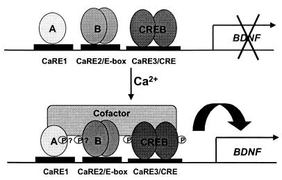
Plasticity is a remarkable feature of the brain, allowing neuronal structure and function to accommodate to patterns of electrical activity. One component of these long-term changes is the activity-driven induction of new gene expression, which is required for both the long-lasting long-term potentiation of synaptic transmission associated with learning and memory, and the activity dependent survival events that help to shape and wire the brain during development. We have characterized molecular mechanisms by which neuronal membrane depolarization and subsequent calcium influx into the cytoplasm lead to the induction of new gene transcription. We have identified three points within this cascade of events where the specificity of genes induced by different types of stimuli can be regulated. By using the induction of the gene that encodes brain-derived neurotrophic factor (BDNF) as a model, we have found that the ability of a calcium influx to induce transcription of this gene is regulated by the route of calcium entry into the cell, by the pattern of phosphorylation induced on the transcription factor cAMP-response element (CRE) binding protein (CREB), and by the complement of active transcription factors recruited to the BDNF promoter. These results refine and expand the working model of activity-induced gene induction in the brain, and help to explain how different types of neuronal stimuli can activate distinct transcriptional responses.


Figures

Figure 1
Figure 1
Calcium-activated signaling pathways that regulate gene transcription. In neurons, neurotransmitter reception and membrane depolarization lead to the opening of ligand- and voltage-gated calcium channels. Subsequent calcium influx across the plasma membrane drives the activation of a number of signaling molecules, including the calcium-sensitive adenylate cyclase, calcium/calmodulin-activated kinases, and Ras. Each of these molecules activates a cascade of signaling proteins that amplifies the calcium signal and carries it to the nucleus. Dashed lines represent the components of each pathway that are proposed to translocate into the nucleus. Nuclear kinases including protein kinase A, CaMK-IV, and members of the Rsk family phosphorylate CREB at Ser-133, rendering it competent to mediate transcription of genes such as BDNF. [Reproduced with permission from ref. (Copyright 1999, Annual Reviews, http://AnnualReviews.org).]
Figure 2
Figure 2
The prevailing view for the mechanism of calcium-dependent CREB activation. CREB sits prebound as a dimer to the CRE in unstimulated cells. In response to neuronal stimulation and the activation of CREB kinases, CREB is phosphorylated at Ser-133 within the kinase-inducible domain (KID), allowing it to bind the KIX domain of CBP (or potentially a CBP dimer) recruiting this coactivator to the promoter. CBP promotes transcriptional activation in part through binding indirectly to the RNA polymerase II via an interaction with the RNA helicase A (RHA) protein, and thus CBP recruits the polymerase complex onto promoters bound by Ser-133-phosphorylated CREB. Other CREB-mediated interactions with the basal transcription machinery may also help to promote transcription. Through a glutamine-rich region (Q2), CREB binds to TAF130, a component of TFIID, and also to TFIIB. These complexes of proteins associate with the TATA binding protein (TBP) at the TATA box that is found just proximal to the initiation site of many genes. [Reproduced with permission from ref. (Copyright 1999, Annual Reviews, http://AnnualReviews.org).]
Figure 3
Figure 3
Calcium-dependent induction of BDNF exon III expression. (A) The BDNF gene has four potential initial exons, each of which can be alternately spliced to a single 3′ exon containing the complete BDNF coding sequence. Each of these splice variants can use one of two alternative polyadenylation sites within the 3′ untranslated region, generating a total of eight distinct transcripts, all of which encode the same protein sequence. (B) Cultured embryonic cortical neurons were depolarized with 50 mM KCl for the times indicated, then RNA was harvested from the cells for quantitative reverse transcription (RT)-PCR analysis by using probes specific for the four initial exons of the BDNF transcripts or for glyceraldehyde-3-phosphate dehydrogenase (GAPDH). Exon III is strongly induced within 180 min of depolarization. GAPDH is constant over the time course examined, serving as a control for RNA input and reverse transcription efficiency. [Reproduced with permission from ref. (Copyright 1998, Elsevier Science).]
Figure 4
Figure 4
Three transcription factors bind to the BDNF promoter III CaREs and coordinately regulate transcription. Three elements within BDNF promoter III are required for calcium-dependent induction of a luciferase reporter gene, and three distinct transcription factors bind to these elements. “A” represents the novel calcium-regulated transcription factor that binds to CaRE1, and “B” represents the basic helix–loop–helix family member that binds the CaRE2/E-box element. CREB binds the CaRE3/CRE. In response to depolarization and the activation of calcium-signaling pathways, all three factors are activated to promote transcription, potentially through the phosphorylation-dependent recruitment of a common transcriptional coactivator.

References

    1. Ghosh A, Greenberg M E. Science. 1995;268:239–247. - PubMed
    1. Franklin J L, Johnson E M. Trends Neurosci. 1992;15:501–508. - PubMed
    1. Lynch G, Larson J, Kelso S, Barrionuevo G, Schottler F. Nature (London) 1983;305:716–721. - PubMed
    1. Wu L, Wells D, Tay J, Mendis D, Abbott M A, Barnitt A, Quinlan E, Heynen A, Fallon J R, Richter J D. Neuron. 1998;21:1129–3119. - PubMed
    1. Aakalu G, Smith W B, Nguyen N, Jiang C, Schuman E M. Neuron. 2001;30:489–502. - PubMed

Publication types

LinkOut - more resources