. 2001 Sep 25;98(20):11062-8.
doi: 10.1073/pnas.191353898.

Regulation of cyclin-dependent kinase 5 and casein kinase 1 by metabotropic glutamate receptors

Affiliations

Regulation of cyclin-dependent kinase 5 and casein kinase 1 by metabotropic glutamate receptors

F Liu et al. Proc Natl Acad Sci U S A. .

Abstract

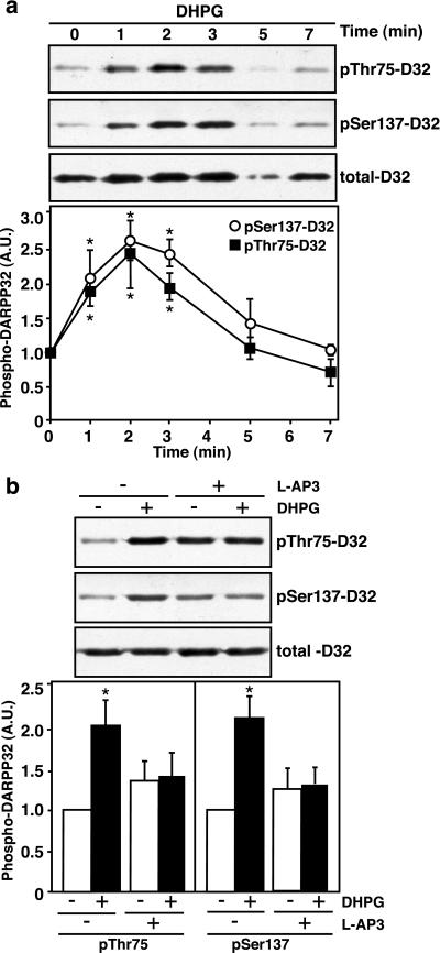
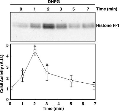
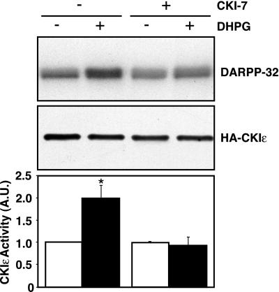
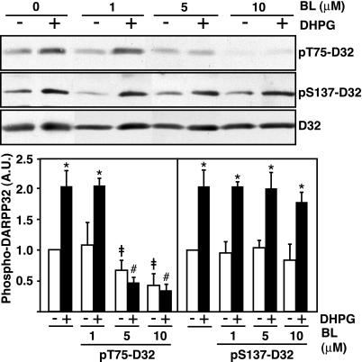
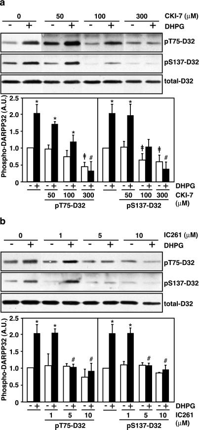
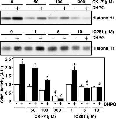
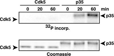
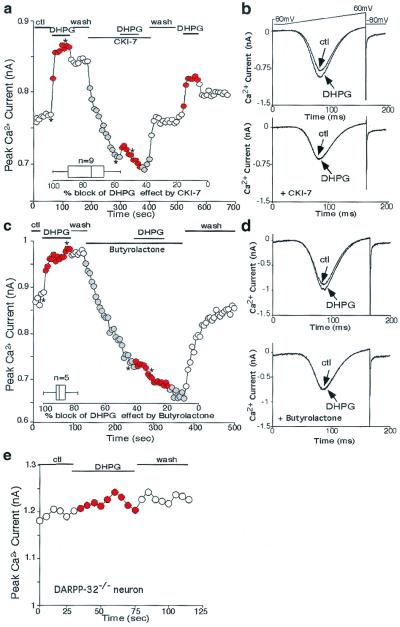
Cyclin-dependent kinase 5 (Cdk5) is a multifunctional neuronal protein kinase that is required for neurite outgrowth and cortical lamination and that plays an important role in dopaminergic signaling in the neostriatum through phosphorylation of Thr-75 of DARPP-32 (dopamine and cAMP-regulated phosphoprotein, molecular mass 32 kDa). Casein kinase 1 (CK1) has been implicated in a variety of cellular functions such as DNA repair, circadian rhythm, and intracellular trafficking. In the neostriatum, CK1 has been found to phosphorylate Ser-137 of DARPP-32. However, first messengers for the regulation of Cdk5 or CK1 have remained unknown. Here we report that both Cdk5 and CK1 are regulated by metabotropic glutamate receptors (mGluRs) in neostriatal neurons. (S)-3,5-dihydroxyphenylglycine (DHPG), an agonist for group I mGluRs, increased Cdk5 and CK1 activities in neostriatal slices, leading to the enhanced phosphorylation of Thr-75 and Ser-137 of DARPP-32, respectively. The effect of DHPG on Thr-75, but not on Ser-137, was blocked by a Cdk5-specific inhibitor, butyrolactone. In contrast, the effects of DHPG on both Thr-75 and Ser-137 were blocked by CK1-7 and IC261, specific inhibitors of CK1, suggesting that activation of Cdk5 by mGluRs requires CK1 activity. In support of this possibility, the DHPG-induced increase in Cdk5 activity, measured in extracts of neostriatal slices, was abolished by CK1-7 and IC261. Treatment of acutely dissociated neurons with DHPG enhanced voltage-dependent Ca(2+) currents. This enhancement was eliminated by either butyrolactone or CK1-7 and was absent in DARPP-32 knockout mice. Together these results indicate that a CK1-Cdk5-DARPP-32 cascade may be involved in the regulation by mGluR agonists of Ca(2+) channels.


Figures

Figure 1
Figure 1
Activation by DHPG of group I mGluRs increases phosphorylation of Thr-75 and Ser-137 on DARPP-32. Mouse neostriatal slices were treated with DHPG for various periods of time as indicated. Slice homogenates were analyzed by SDS/PAGE and immunoblotting with phospho-Thr-75, phospho-Ser-137, and total DARPP-32 antibodies. (a) Time course of treatment with DPHG (100 μM). (b) Slices were preincubated without or with the group I mGluR antagonist L-AP3 (100 μM) for 20 min before treatment with DHPG for 2 min. Cumulative data (means ± SEM) obtained from five experiments are shown in a and b Lower. Data were normalized to values for untreated slices. *, P < 0.05, Student's t test, compared with untreated slices.
Figure 2
Figure 2
Activation of mGluR1s transiently increases Cdk5 activity. Mouse neostriatal slices were treated with DHPG for various periods as indicated. Slices were homogenized and Cdk5 was immunoprecipitated by using anti-Cdk5 (C-8) antibody. Cdk5 activity was assayed by using histone H-1 as substrate, and samples were analyzed by SDS/PAGE and autoradiography. (Upper) An autoradiogram of histone H-1 phosphorylation. (Lower) Cumulative data. The autoradiograms were analyzed by using a PhosphoImager. Data for three experiments (means ± SEM) were normalized to the values obtained at 0 min. *, P < 0.05, Student's t test, compared with 0 min.
Figure 3
Figure 3
Activation of mGluR1s stimulates CK1 activity. N2a cells were transiently transfected with HA-tagged CK1ɛ. Cells were preincubated without or with the CK1 inhibitor CK1–7 (100 μM) for 30 min before treatment with DHPG (100 μM) for an additional 2 min. HA-CK1ɛ was immunoprecipitated and CK1 was assayed by using DARPP-32 as a substrate. Samples were analyzed by SDS/PAGE and autoradiography. (Top) Autoradiogram of DARPP-32 phosphorylation. (Middle) Immunoblot of the level of expression of HA-CK1ɛ. (Bottom) Cumulative data of kinase activity obtained from five experiments (means ± SEM). Data were normalized to values obtained for untreated cells. *, P < 0.05, Student's t test, compared with untreated cells.
Figure 4
Figure 4
A Cdk5 inhibitor, butyrolactone, blocks the effect of DHPG on phosphorylation of DARPP-32 on Thr-75 (Cdk5 site) but not on Ser-137 (CK1 site). Mouse neostriatal slices were preincubated with 0, 1, 5, or 10 μM butyrolactone (BL) for 30 min, and then DHPG (100 μM) was added for an additional 2 min. Slice homogenates were analyzed by SDS/PAGE and immunoblotting with phospho-Thr-75, phospho-Ser-137, and total DARPP-32 antibodies. (Upper) Immunoblots. (Lower) Cumulative data (means ± SEM) obtained from three experiments. Data were normalized to values obtained for untreated slices. *, P < 0.05, Student's t test, compared with slices in the absence of DHPG; ‡, P < 0.05, Student's t test, compared with untreated slices; #, P < 0.05, Student's t test, compared with slices treated with DHPG alone.
Figure 5
Figure 5
CK1 inhibitors, CK1–7 and IC261, block the effects of DHPG on phosphorylation of DARPP-32 on both Ser-137 (CK1 site) and Thr-75 (Cdk5 site). Mouse neostriatal slices were preincubated for 30 min with (a) 0, 50, 100, or 300 μM CK1–7 or (b) 0, 1, 5, or 10 μM IC261, and then DHPG (100 μM) was added for an additional 2 min. Slice homogenates were analyzed by SDS/PAGE and immunobloting with phospho-Ser-137, phospho-Thr-75, and total DARPP-32 antibodies. (Upper) Immunoblots. (Lower) Cumulative data (means ± SEM) obtained from three experiments. Data were normalized to values obtained for untreated slices. *, P < 0.05, Student's t test, compared with slices in the absence of DHPG; ‡, P < 0.05, Student's t test, compared with untreated slices; #, P < 0.05, Student's t test, compared with slices treated with DHPG alone.
Figure 6
Figure 6
CK1 inhibitors block the effect of DHPG on Cdk5 activity. Mouse neostriatal slices were preincubated with 0, 50, 100, or 300 μM CK1–7 or 0, 1, 5, or 10 μM IC261 for 30 min, and then DHPG (100 μM) was added for an additional 2 min. Slices were homogenized and Cdk5 was immunoprecipitated with anti-Cdk5 (C-8) antibody. Cdk5 activity was assayed by using histone H-1 as substrate and samples were analyzed by SDS/PAGE and autoradiography. (Upper) Autoradiograms indicating histone H-1 phosphorylation. (Lower) Cumulative data (means ± SEM) from three experiments. The autoradiograms were analyzed by using a PhosphorImager, and the data were normalized to values obtained for untreated slices. *, P < 0.05, Student's t test, compared with slices in the absence of DHPG; ‡, P < 0.05, Student's t test, compared with untreated slices; #, P < 0.05, Student's t test, compared with slices treated with DHPG alone.
Figure 7
Figure 7
CK1 phosphorylates p35 in vitro. Cdk5 (Left) or p35 (Right) was incubated in vitro with CK1 and [32P]ATP for various times as indicated. Samples were analyzed by SDS/PAGE and autoradiography. 32P incorporation is shown in the autoradiogram (Upper). Total amounts of Cdk5 and p35 are shown in the Coomassie-stained gel (Lower).
Figure 8
Figure 8
Activation by DHPG of group I mGluRs enhances Ca2+ current, and this enhancement is blocked by inhibiting CK1 or cdk5 and is absent in DARPP-32 knockout mice. (a and c) Peak Ca2+ current evoked by a voltage ramp protocol as a function of time and drug application. (a) DHPG (100 μM) increased Ca2+ current. In the presence of CK1–7 (100 μM), basal Ca2+ currents were reduced and the DHPG effect was attenuated. Washing out CK1–7 led to recovery of the DHPG effect. (c) DHPG (100 μM) increased Ca2+ current. In the presence of butyrolactone (25 μM), basal Ca2+ currents were reduced and the DHPG effect was eliminated. (b and d) Representative current traces showing the modulation by DHPG before and after the application of (b) CK1–7 or (d) butyrolactone (at time points denoted by *). (Insets) Box plot summaries showing the percentage block of the DHPG effect on Ca2+ currents by (a) CK1–7 and (c) butyrolactone. (e) Plot of peak Ca2+ current as a function of time and agonist application in a representative neostriatal neuron from a DARPP-32 knockout mouse. DHPG (100 μM) had no effect on Ca2+ current in neurons from DARPP-32−/− mice (n = 9).

References

    1. Tsai L H, Takahashi T, Caviness V S, Jr, Harlow E. Development (Cambridge, UK) 1993;119:1029–1040. - PubMed
    1. Lew J, Huang Q Q, Qi Z, Winkfein R J, Aebersold R, Hunt T, Wang J H. Nature (London) 1994;371:423–426. - PubMed
    1. Tsai L H, Delalle I, Caviness V S, Jr, Chae T, Harlow E. Nature (London) 1994;371:419–423. - PubMed
    1. Ohshima T, Ward J M, Huh C G, Longenecker G, Veeranna, Pant H C, Brady R O, Martin L J, Kulkarni A B. Proc Natl Acad Sci USA. 1996;93:11173–11178. - PMC - PubMed
    1. Chae T, Kwon Y T, Bronson R, Dikkes P, Li E, Tsai L H. Neuron. 1997;18:29–42. - PubMed

Publication types

MeSH terms