Comparative Study
. 2001 Sep 25;98(20):11236-41.
doi: 10.1073/pnas.201373498.

Purification and characterization of human DNA damage checkpoint Rad complexes

Affiliations
Comparative Study

Purification and characterization of human DNA damage checkpoint Rad complexes

L A Lindsey-Boltz et al. Proc Natl Acad Sci U S A. .

Abstract

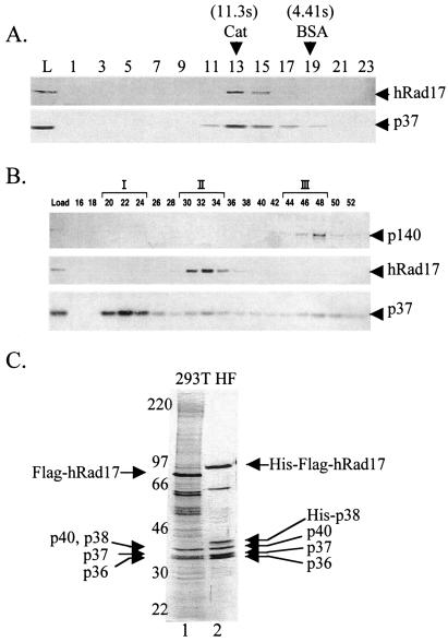
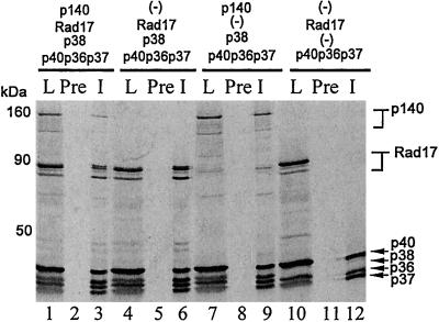
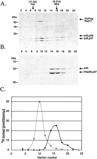
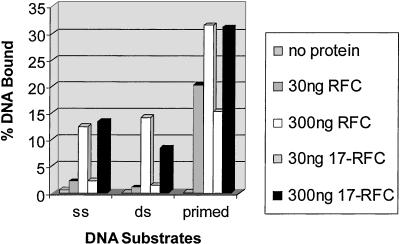
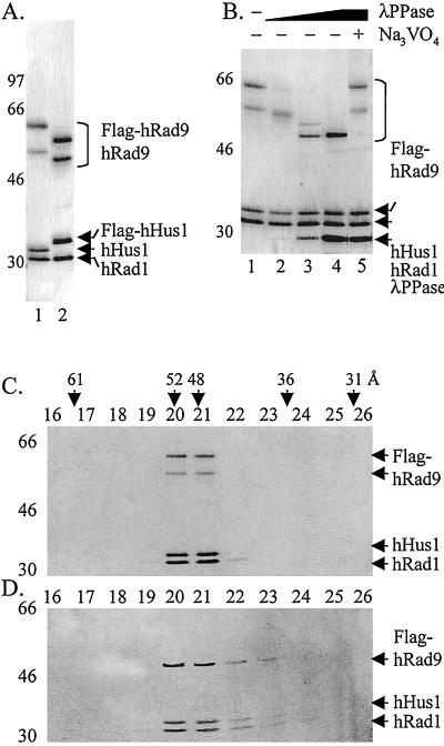
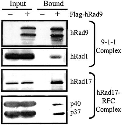
Checkpoint Rad proteins function early in the DNA damage checkpoint signaling cascade to arrest cell cycle progression in response to DNA damage. This checkpoint ensures the transmission of an intact genetic complement to daughter cells. To learn about the damage sensor function of the human checkpoint Rad proteins, we purified a heteropentameric complex composed of hRad17-RFCp36-RFCp37-RFCp38-RFCp40 (hRad17-RFC) and a heterotrimeric complex composed of hRad9-hHus1-hRad1 (checkpoint 9-1-1 complex). hRad17-RFC binds to DNA, with a preference for primed DNA and possesses weak ATPase activity that is stimulated by primed DNA and single-stranded DNA. hRad17-RFC forms a complex with the 9-1-1 heterotrimer reminiscent of the replication factor C/proliferating cell nuclear antigen clamp loader/sliding clamp complex of the replication machinery. These findings constitute biochemical support for models regarding the roles of checkpoint Rads as damage sensors in the DNA damage checkpoint response of human cells.


Figures

Figure 1
Figure 1
Isolation of hRad17-RFC. (A) Glycerol gradient sedimentation of hRad17 present in crude HeLa extracts. HeLa CFE was subjected to glycerol gradient centrifugation as described in Materials and Methods. Fractions were collected from the bottom of the tube and analyzed by Western blotting with anti-hRad17 and anti-RFCp37 antibodies. The sedimentation position of reference proteins in a parallel gradient is indicated. L, load. (B) Separation of at least three forms of RFC. Fractions from the SP Sepharose column (see Materials and Methods) were analyzed by Western blotting by using the indicated antibodies. The three RFC complexes are represented by I, II, and III. (C) Purification the hRad17-RFC complex from transfected human 293T cells and baculovirus-infected insect HF cells. 293T cells were transfected with pcDNA4-Flag-hRad17 and purified by using anti-Flag agarose (lane 1). The hRad17-RFC complex was reconstituted in insect cells by coinfection with five recombinant viruses capable of expressing His6-Flag-hRad17 and each RFC small subunit (lane 2). The p38 subunit contains a His6 tag. The complex was purified by chromatography with Ni-NTA and then anti-Flag agarose as described in Materials and Methods. The proteins were visualized after SDS/PAGE by silver staining. The amounts of protein loaded in lanes 1 and 2 were 1 and 0.5 μg, respectively.
Figure 2
Figure 2
Order of assembly of hRad17-RFC. An in vitro transcription/translation system containing the indicated expression vectors was used to radiolabel the corresponding proteins. The products were immunoprecipitated with anti-p37 antibodies, separated on SDS/PAGE, and analyzed by autoradiography. L, load; Pre, preimmune serum, and I, immune (anti-p37) serum.
Figure 3
Figure 3
The copurification of ATPase activity with hRad17-RFC. (A and B) Analysis of glycerol gradient fractions of hRad17-RFC and p36-p37-p40 core RFC by SDS/PAGE and Coomassie blue staining. hRad17-RFC (0.2 ml, 0.73 mg/ml) or the p36-p37-p40 core RFC complex (0.2 ml, 1 mg/ml) was loaded onto a 5-ml 15–35% glycerol gradient and centrifuged for 20 h at 4° at 250,000 × g. Fractions were collected from the bottom of the gradients and 10 μl of each was loaded onto the gels. (C) ATPase activity of hRad17-RFC and core RFC. Each fraction (1 μl) was analyzed for ATPase activity in the presence of 12.5 μM (as nucleotides) poly dA4000:oligo dT12–18 as indicated in Materials and Methods. Peak fractions, fraction 10 for hRad17-RFC and fraction 16 for core RFC, contained 25 and 59 ng of protein/μl, respectively.
Figure 4
Figure 4
DNA-binding activity of hRad17-RFC. The indicated amounts of RFC or hRad17-RFC were incubated in a reaction mixture (30 μl) containing either 40 fmol of 32P-labeled 50-nt single-stranded (ss), 50-bp dsDNA or a primed 50-bp dsDNA with a 50-nt ssDNA (5′ overhang) in 25 mM Hepes (pH 7.5), 175 mM NaCl, 3 mM MgCl2, 1 mM DTT, and 0.1 mg/ml BSA for 30 min on ice. The fraction of DNA bound to the protein was quantitated by a nitrocellulose filter-binding assay. The details of the DNA substrates used were described (28).
Figure 5
Figure 5
Purification and characterization of the checkpoint 9-1-1 complex. (A) Purification of the 9-1-1 complex. HF insect cells were infected with baculoviruses expressing all three subunits with tags where indicated, and the complex was purified by immunoaffinity chromatography as described in Materials and Methods. Lane 1, complex purified through the Flag tag on hRad9 (0.4 μg loaded) and lane 2, complex purified through the Flag tag on hHus1 (0.4 μg loaded). (B) Effect of λ phosphatase on the 9-1-1 complex. The complex was treated with 0, 3.2, 16, 80, and 80 units of phosphatase in lanes 1–5, respectively, under conditions described by the manufacturer. After 1-h incubation at 30°C, the products were analyzed by SDS/PAGE and silver staining. (C and D) Dephosphorylated hRad9 remains in the 9-1-1 complex. Checkpoint 9-1-1 complexes, before (C) or after (D) λ phosphatase treatment, were separated by gel filtration, and the fractions were analyzed by SDS/PAGE and silver staining.
Figure 6
Figure 6
Isolation of the checkpoint Rad complex. HF cells infected with either all of the subunits of the hRad17-RFC and 9-1-1 complexes, including Flag-tagged hRad9, or all of the subunits except hRad9 were lysed, and the complex was purified by immunoaffinity chromatography and analyzed by Western blotting as described in Materials and Methods.

References

    1. Hartwell L H, Weinert T A. Science. 1989;246:629–634. - PubMed
    1. Lowndes N F, Murguia J R. Curr Opin Genet Dev. 2000;10:17–25. - PubMed
    1. O'Connell M J, Walworth N C, Carr A M. Trends Cell Biol. 2000;10:296–303. - PubMed
    1. Zhou B B, Elledge S J. Nature (London) 2000;408:433–439. - PubMed
    1. Rhind N, Russell P. Curr Opin Cell Biol. 1998;10:749–758. - PMC - PubMed

Publication types

MeSH terms