. 2001 Sep 25;98(20):11417-22.
doi: 10.1073/pnas.191175898.

WWOX: a candidate tumor suppressor gene involved in multiple tumor types

Affiliations

WWOX: a candidate tumor suppressor gene involved in multiple tumor types

A J Paige et al. Proc Natl Acad Sci U S A. .

Abstract

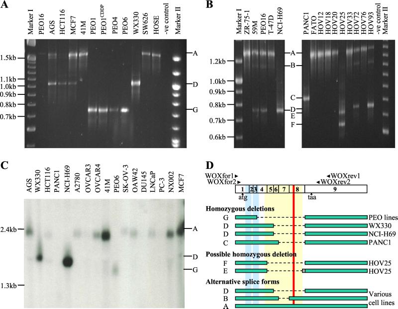
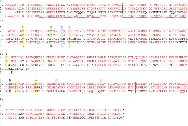
We previously reported the construction of a P1-derived artificial chromosome (PAC) contig encompassing a set of homozygous deletions of chromosome 16q23-24.1 found in primary ovarian tumor material and several tumor cell lines. Using these PAC clones in a cDNA selection experiment, we have isolated a Sau3A fragment homologous to the WWOX transcript (GenBank accession no. ) from normal human ovarian surface epithelial (HOSE) cells. We demonstrate the homozygous deletion of WWOX exons from ovarian cancer cells and three different tumor cell lines. We also identify an internally deleted WWOX transcript from a further primary ovarian tumor. In three of these samples the deletions result in frameshifts, and in each case the resulting WWOX transcripts lack part, or all, of the short chain dehydrogenase domain and the putative mitochondrial localization signal. Sequencing revealed several missense polymorphisms in tumor cell lines and identified a high level of single nucleotide polymorphism (SNP) within the WWOX gene. This evidence strengthens the case for WWOX as a tumor suppressor gene in ovarian cancer and other tumor types.


Figures

Figure 1
Figure 1
(A) An example RT-PCR from tumor cell line RNA using WOXfor1 and WOXrev1 primers. PCR products were separated on 2% Metaphor agarose (Cambrex, East Rutherford, NJ) in 1× 0.04 M Tris-acetate/0.001 M EDTA (TAE) at 70 V for 4 h. The expected 1.56-kb product representing normal FORII (transcript A) can be seen in several cell lines and in HOSE. Aberrant transcripts D and G can be seen in the homozygously deleted WX330 and PEO6 cell lines, respectively. The PEO1CDDP, PEO1, and PEO4 cell lines were derived at different time points from the same ovarian cancer patient as the PEO6 cell line. All four of these lines exhibit homozygous deletion of 16q23 and produce the aberrant transcript G. Several cell lines (e.g., MCF7) show both transcript A and transcript D. (B) An example nested RT-PCR from tumor cell line and primary ovarian tumor (HOV) RNA using WOXfor2 and WOXrev2 primers. PCR products were separated on 2% Metaphor agarose in 1× TAE at 70 V for 4 h. The expected 1.29-kb product representing normal FORII (transcript A) can be seen in several lanes. Aberrant transcripts C and D can be seen in the homozygously deleted PANC1 and NCI-H69 cell lines, respectively. Two aberrant transcripts (E and F) were detected in the primary ovarian tumor sample HOV25. Several samples exhibited both transcript A and additional products B and D. (C) Northern Blot of tumor cell lines hybridized with a WWOX coding region probe. Aberrant, short transcripts were detected from WX330 and NCI-H69, and from PEO6, corresponding to the predicted sizes of transcripts D and G, respectively—thus confirming the homozygous loss of WWOX exons in these samples. No signal was detected from PANC1. Transcript A was found to be the predominant WWOX splice form in the remaining cell lines. (D) Schematic representation of the different transcripts identified by RT-PCR. Positions of PCR primers used for primary and nested RT-PCR are shown. White boxes indicate the nine exons of the WWOX (FORII) transcript, blue boxes show the position of the WW domains, a yellow box indicates the adh domain, and a red line represents the putative substrate-binding site. Each transcript is shown by a green bar with the dashed line representing the deleted portion of the transcript. The gray box in transcript E represents the 76-bp insertion (GenBank accession no. AF325433). Aberrant transcripts C, E, F, and G are out-of-frame deletions, whereas transcript D exhibits an in-frame deletion. However, in every case, the substrate-binding site, putative mitochondrial localization signal, and most of the adh domain are deleted.
Figure 2
Figure 2
Polymorphisms within the coding region of the WWOX gene. CLUSTALX alignment of WWOX protein sequences showing amino acid residues altered by nucleotide changes identified through mutation screening. Line 1: Polymorphic residues in Human WWOX (FORII isoform) corresponding to polymorphisms listed in Table 1. Line 2: Human WWOX (FORII isoform). Line 3: Mouse Wox1. Line 4: Drosophila Wwox (GenPept accession no. AAF52587). Line 5: Human WWOX (FORIII isoform). Line 6: Polymorphic residues in Human WWOX (FORIII isoform) corresponding to polymorphisms listed in Table 1. Amino acids indicated in red show conservation with the Human WWOX FORII isoform (Line 2). Blue boxes show silent changes where the amino acid is unaltered by the nucleotide change. Yellow boxes indicate polymorphic residues due to SNPs. Green boxes indicate missense polymorphisms detected only in tumor cell lines or normal tissue from cancer patients. The WW domains are shown as dashed boxes and the adh domain as a solid box.

References

    1. Chen T, Sahin A, Aldaz C M. Cancer Res. 1996;56:5605–5609. - PubMed
    1. Paris P L, Witte J S, Kupelian P A, Levin H, Klein E A, Catalona W J, Casey G. Cancer Res. 2000;60:3645–3649. - PubMed
    1. Piao Z, Park C, Kim J J, Kim H. Br J Cancer. 1999;80:850–854. - PMC - PubMed
    1. Paige A J W, Taylor K J, Stewart A, Sgouros J G, Gabra H, Sellar G C, Smyth J F, Porteous D J, Watson J E V. Cancer Res. 2000;60:1690–1697. - PubMed
    1. Mangelsdorf M, Ried K, Woollatt E, Dayan S, Eyre H, Finnis M, Hobson L, Nancarrow J, Venter D, Baker E, Richards R I. Cancer Res. 2000;60:1683–1689. - PubMed

Publication types

MeSH terms