. 2001 Sep 25;98(20):11621-6.
doi: 10.1073/pnas.191384198.

Fruiting body formation by Bacillus subtilis

Affiliations

Fruiting body formation by Bacillus subtilis

S S Branda et al. Proc Natl Acad Sci U S A. .

Abstract

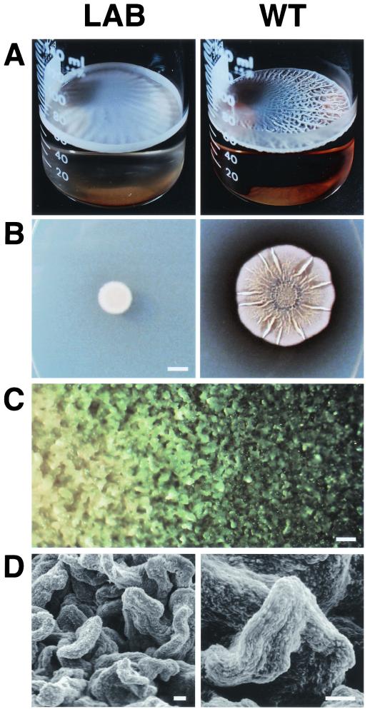
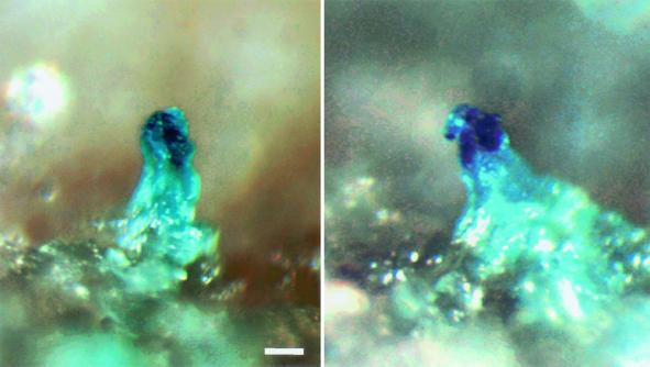
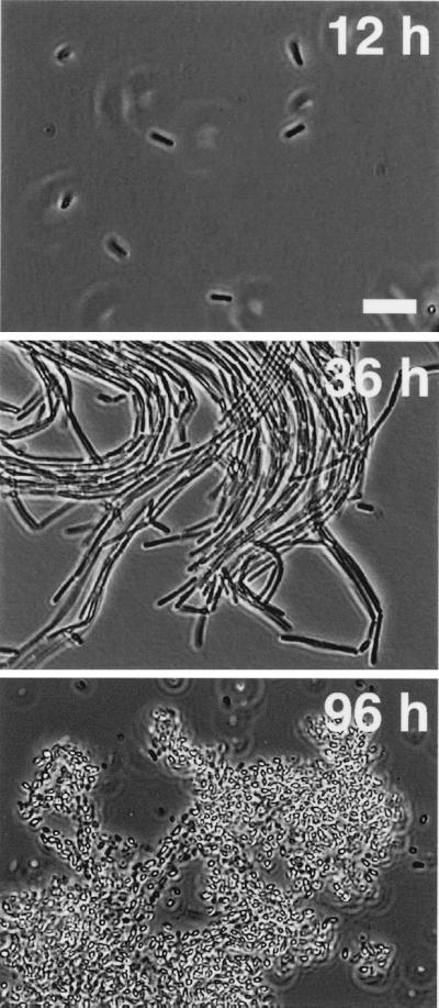
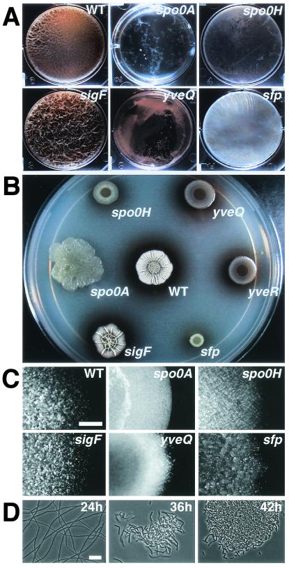
Spore formation by the bacterium Bacillus subtilis has long been studied as a model for cellular differentiation, but predominantly as a single cell. When analyzed within the context of highly structured, surface-associated communities (biofilms), spore formation was discovered to have heretofore unsuspected spatial organization. Initially, motile cells differentiated into aligned chains of attached cells that eventually produced aerial structures, or fruiting bodies, that served as preferential sites for sporulation. Fruiting body formation depended on regulatory genes required early in sporulation and on genes evidently needed for exopolysaccharide and surfactin production. The formation of aerial structures was robust in natural isolates but not in laboratory strains, an indication that multicellularity has been lost during domestication of B. subtilis. Other microbial differentiation processes long thought to involve only single cells could display the spatial organization characteristic of multicellular organisms when studied with recent natural isolates.


Figures

Figure 1
Figure 1
Architecture of B. subtilis pellicles and colonies. (A) LAB and WT pellicles. Overnight cultures were diluted 1,000-fold into MSgg medium, and 60 ml was transferred to 150-ml Pyrex beakers. These cultures were incubated at 25°C without agitation for 5 days and then photographed. (B) LAB and WT colonies. Five microliters from overnight cultures were spotted onto a dry minimal agar plate. The plate was incubated at 25°C for 5 days and then photographed. (Bar = 5 mm.) (C) Close-up of the edge of a WT colony. A colony was grown at 25°C for 2 days and then photographed at 16× magnification with a dissection microscope equipped with a charge-coupled device video camera. (Bar = 100 μm.) (D) Scanning electron micrographs of a WT colony. A colony similar to that shown in C was photographed at 600× magnification (Left) and 1,000× magnification (Right). (Bars = 10 μm.)
Figure 2
Figure 2
Sporulation sites on the surfaces of WT colonies. WT(sspE-lacZ) bacteria were streaked on a minimal agar plate containing X-Gal (300 μg/ml), incubated at 25°C for 3 days, and then photographed at 130× magnification as described for Fig 1C. (Bar = 50 μm.)
Figure 3
Figure 3
B. subtilis pellicle development at the cellular level. WT cultures similar to those shown in Fig. 1A were incubated at 25°C without agitation. At the times indicated, samples were withdrawn from the air–medium interface and examined at 600× magnification with a phase-contrast microscope equipped with a charge-coupled device video camera. (Bar = 5 μm.)
Figure 4
Figure 4
Developmental properties of various B. subtilis mutants. Starting cultures were grown as described in the legend of Fig. 1; see text for description of each mutant. (A) Mutant pellicles. All starting cultures were diluted 1,000-fold, and 12 ml was transferred to the wells of 6-well microtiter plates. These cultures were incubated and photographed as described for Fig. 1A. (B) Mutant colonies at low magnification. Five microliters from each starting culture was spotted on an MSgg agar plate, and the plate was incubated and photographed as described for Fig. 1B. (C) Mutant colonies at high magnification. Colonies were incubated and photographed as described in Fig. 1C. (Bar = 0.5 μm.) (D) Development of yveQ mutant pellicles at the cellular level. Cultures similar to those of A were incubated at 25°C without agitation; at the times indicated, samples were withdrawn from the air–medium interface and examined by phase-contrast microscopy as described in the legend for Fig. 3.

References

    1. Losick R, Shapiro L, editors. Microbial Development. Plainview, NY: Cold Spring Harbor Lab. Press; 1984.
    1. Dworkin M. Developmental Biology of the Bacteria. San Francisco: Benjamin/Cummings Science; 1985.
    1. Brun Y V, Shimkets L J, editors. Prokaryotic Development. Washington, DC: Am. Soc. Microbiol.; 2000.
    1. Piggot, P. & Losick, R. (in press) in Bacillus subtilis and Its Closest Relatives: From Genes to Cells, eds. Sonenshein, A. L., Hoch, J. A. & Losick, R. (Am. Soc. Microbiol., Washington, DC), in press.
    1. Setlow, P. (in press) in Bacillus subtilis and Its Closest Relatives: From Genes to Cells, eds. Sonenshein, A. L., Hoch, J. A. & Losick, R. (Am. Soc. Microbiol., Washington, DC), in press.

Publication types

MeSH terms

Substances