. 2001 Sep 25;98(20):11627-32.
doi: 10.1073/pnas.191384398.

A universal protein-protein interaction motif in the eubacterial DNA replication and repair systems

Affiliations

A universal protein-protein interaction motif in the eubacterial DNA replication and repair systems

B P Dalrymple et al. Proc Natl Acad Sci U S A. .

Abstract

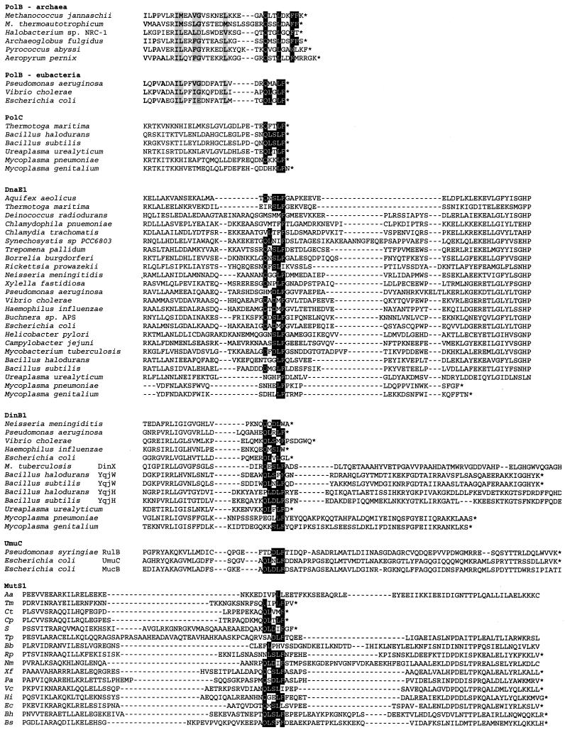
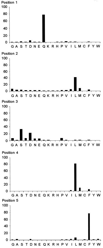
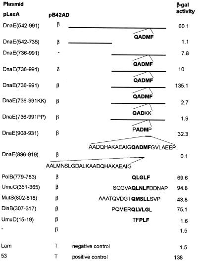
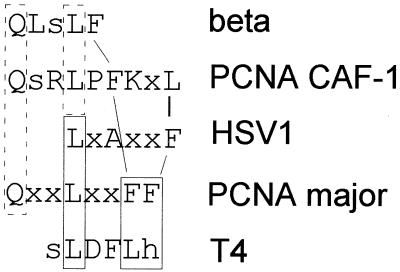
The interaction between DNA polymerases and sliding clamp proteins confers processivity in DNA synthesis. This interaction is critical for most DNA replication machines from viruses and prokaryotes to higher eukaryotes. The clamp proteins also participate in a variety of dynamic and competing protein-protein interactions. However, clamp-protein binding sequences have not so far been identified in the eubacteria. Here we show from three lines of evidence, bioinformatics, yeast two-hybrid analysis, and inhibition of protein-protein interaction by modified peptides, that variants of a pentapeptide motif (consensus QL[SD]LF) are sufficient to enable interaction of a number of proteins with an archetypal eubacterial sliding clamp (the beta subunit of Escherichia coli DNA polymerase III holoenzyme). Representatives of this motif are present in most sequenced members of the eubacterial DnaE, PolC, PolB, DinB, and UmuC families of DNA polymerases and the MutS1 mismatch repair protein family. The component tripeptide DLF inhibits the binding of the alpha (DnaE) subunit of E. coli DNA polymerase III to beta at microM concentration, identifying key residues. Comparison of the eubacterial, eukaryotic, and archaeal sliding clamp binding motifs suggests that the basic interactions have been conserved across the evolutionary landscape.


Figures

Figure 1
Figure 1
Alignment of the regions containing the putative β-binding peptides from members of the eubacterial PolB, PolC, DnaE1, DinB1, UmuC, and MutS1 families and the PCNA-binding motif in members of the archaeal PolB family. Representative sequences from fully sequenced genomes, including conserved flanking regions, are shown. Amino acids shown as white with black backgrounds are matches to the β-binding site consensus peptide sequence, amino acids shown with pale gray backgrounds are conservative substitutions in the archaeal and eubacterial PolB sequences. * indicates the terminal amino acid in the protein sequence. For sources of sequences, see Table 3, which is published as supporting information on the PNAS web site.
Figure 2
Figure 2
Distribution of amino acids in putative β-binding peptides. A single peptide sequence with three or more matches to the motif Qxshh (where x is any amino acid, s is any small amino acid, and h is any hydrophobic amino acid) in the appropriate region of the protein from each member of the PolC (22 examples), PolB (15 examples), DnaE1 (72 examples), UmuC (20 examples), DinB1 (62 examples), and MutS1 (59 examples) families of proteins was included in the analysis. Frequency (%) is plotted (as ordinate) for each amino acid at each position of the pentapeptide motif. For a list of sequences used in the analysis see Table 4, which is published as supporting information on the PNAS web site.
Figure 3
Figure 3
Yeast two-hybrid analysis of β-binding sites. Numbers in brackets in the plasmid names indicate the amino acid range included in the LexA fusion proteins. Plasmid pLexADnaE(542–991) contains the region of dnaE previously identified to contain the β-binding site (19). Plasmid pLexADnaE(542–735) is predicted not to bind to β, based on Kim and McHenry (19). Plasmid pLexA-53 encodes LexA fused to murine p53 protein, pB42AD-T encodes a B42AD domain fused to the simian virus 40 large T antigen, and pLexA-Lam encodes LexA fused to human lamin C.
Figure 4
Figure 4
Comparison of the core sliding clamp-binding motifs. CAF-1 data are from Moggs et al. (34) and additional analysis (data not shown), HSV1 data are from Zuccola et al. (35), and T4 data are from Berdis et al. (33) and Wong and Geiduschek (36). Amino acids for which there is structural data supporting similar roles (35, 37) are in solid boxes; amino acids conserved in position and therefore likely to have similar roles are in dashed boxes; hydrophobic amino acids that are not conserved in position but that contribute significantly to the binding of the peptides are connected by lines.

References

    1. Bruck I, O'Donnell M. J Biol Chem. 2000;275:28971–28983. - PubMed
    1. Fay P J, Johanson K O, McHenry C S, Bambara R A. J Biol Chem. 1981;256:976–983. - PubMed
    1. Kong X P, Onrust R, O'Donnell M, Kuriyan J. Cell. 1992;69:425–437. - PubMed
    1. Rangarajan S, Woodgate R, Goodman M F. Proc Natl Acad Sci USA. 1999;96:9224–9229. - PMC - PubMed
    1. Hughes A J, Jr, Bryan S K, Chen H, Moses R E, McHenry C S. J Biol Chem. 1991;266:4568–4573. - PubMed

Publication types

MeSH terms

LinkOut - more resources