. 2001 Sep 25;98(20):11650-5.
doi: 10.1073/pnas.201243998.

Mapping adolescent brain change reveals dynamic wave of accelerated gray matter loss in very early-onset schizophrenia

Affiliations

Mapping adolescent brain change reveals dynamic wave of accelerated gray matter loss in very early-onset schizophrenia

P M Thompson et al. Proc Natl Acad Sci U S A. .

Abstract

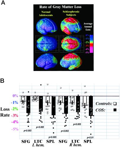
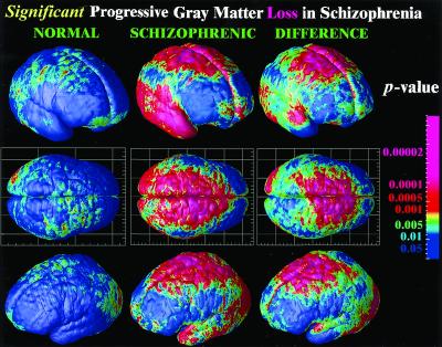
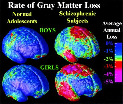
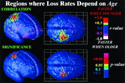
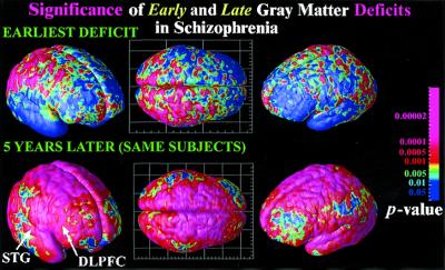
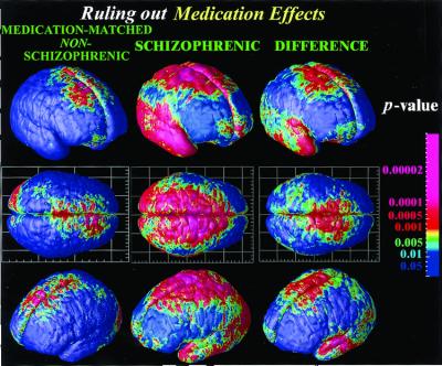
Neurodevelopmental models for the pathology of schizophrenia propose both polygenetic and environmental risks, as well as early (pre/perinatal) and late (usually adolescent) developmental brain abnormalities. With the use of brain mapping algorithms, we detected striking anatomical profiles of accelerated gray matter loss in very early-onset schizophrenia; surprisingly, deficits moved in a dynamic pattern, enveloping increasing amounts of cortex throughout adolescence. Early-onset patients were rescanned prospectively with MRI, at 2-year intervals at three time points, to uncover the dynamics and timing of disease progression during adolescence. The earliest deficits were found in parietal brain regions, supporting visuospatial and associative thinking, where adult deficits are known to be mediated by environmental (nongenetic) factors. Over 5 years, these deficits progressed anteriorly into temporal lobes, engulfing sensorimotor and dorsolateral prefrontal cortices, and frontal eye fields. These emerging patterns correlated with psychotic symptom severity and mirrored the neuromotor, auditory, visual search, and frontal executive impairments in the disease. In temporal regions, gray matter loss was completely absent early in the disease but became pervasive later. Only the latest changes included dorsolateral prefrontal cortex and superior temporal gyri, deficit regions found consistently in adult studies. These emerging dynamic patterns were (i) controlled for medication and IQ effects, (ii) replicated in independent groups of males and females, and (iii) charted in individuals and groups. The resulting mapping strategy reveals a shifting pattern of tissue loss in schizophrenia. Aspects of the anatomy and dynamics of disease are uncovered, in a changing profile that implicates genetic and nongenetic patterns of deficits.


Figures

Figure 1
Figure 1
Average rates of gray matter loss in normal adolescents and in schizophrenia. (A) Three-dimensional maps of brain changes, derived from high-resolution magnetic resonance images (MRI scans) acquired repeatedly from the same subjects, reveal profound, progressive gray matter loss in schizophrenia (Right). Average rates of gray matter loss from 13 to 18 years of age are displayed on average cortical models for the group. Severe loss is observed (red and pink; up to 5% annually) in parietal, motor, and temporal cortices, whereas inferior frontal cortices remain stable (blue; 0–1% loss). Dynamic loss is also observed in the parietal cortices of normal adolescents, but at a much slower rate. (B) Average gray matter loss rates were computed for all 24 subjects in superior frontal gyri (SFG), lateral temporal cortices (LTC), and superior parietal lobules (SPL) in both brain hemispheres. Error bars indicate the standard error of the sample means, by region, in controls and patients. Individual loss rates (in percent per year) are plotted (■, patients; □, controls), showing significant group separation, despite some outliers.
Figure 2
Figure 2
Significance of dynamic gray matter loss in normal adolescents and in schizophrenia. Highly significant progressive loss occurs in schizophrenia in parietal, motor, supplementary motor, and superior frontal cortices. Broad regions of temporal cortex, including the superior temporal gyrus, experience severe gray matter attrition. By comparison of the average rates of loss in disease (middle column) with the loss pattern in normal adolescents (first column), the normal variability in these changes can also be taken into account, and the significance of disease-specific change can be established (last column).
Figure 3
Figure 3
Dynamic changes in male and female teenagers with schizophrenia. A consistent pattern of progressive gray matter loss, in parietal, frontal, and temporal cortices, is observed in independent groups of males and female patients. A single pattern is observable in both boys and girls, supporting the anatomical specificity of the findings.
Figure 4
Figure 4
Mapping nonlinear brain changes and age effects. Dependencies between the rate of gray matter loss and the patient's age are mapped locally and visualized. Parietal regions lose gray matter faster in younger patients (red; r > 0.8; P < 0.001), consistent with an earlier timing of deficits and a slowing of the rates of progression as adolescence continues.
Figure 5
Figure 5
Mapping early and late deficits in schizophrenia. Deficits occurring during the development of schizophrenia are detected by comparing average profiles of gray matter between patients and controls at their first scan (age 13; Upper) and their last scan 5 years later (age 18; Lower). Although severe parietal, motor, and diffuse frontal loss has already occurred (Upper) and subsequently continues (Figs. 1 and 2), the temporal and dorsolateral prefrontal loss characteristic of adult schizophrenia is not found until later in adolescence (Lower), where a process of fast attrition occurs over the next 5 years. The color code shows the significance of these effects.
Figure 6
Figure 6
Mapping brain change in medication-matched subjects not satisfying criteria for schizophrenia. No temporal lobe deficits are found, suggesting that the progression of the deficits into the temporal lobe is specific to schizophrenia, regardless of medication (and regardless of gender; Fig. 3). Nonetheless, these patients share some symptoms with schizophrenics, exhibiting frontal deficits in a similar anatomical pattern. These frontal deficits (i.e., gray matter loss rates) are statistically significant relative to healthy controls but significantly smaller in magnitude than the greatly accelerated loss rates in schizophrenia.

References

    1. Jacobsen L K, Rapoport J L. J Child Psychol Psychiatry. 1998;39:101–113. - PubMed
    1. Feinberg I. J Psychiatr Res. 1982;17:319–334. - PubMed
    1. Weinberger D R. In: Schizophrenia. Hirsch S R, Weinberger D R, editors. London: Blackwood; 1995. pp. 294–323.
    1. Barondes S H, Alberts B M, Andreasen N C, Bargmann C, Benes F, Goldman-Rakic P, Gottesman I, Heinemann S F, Jones E G, Kirschner M, et al. Proc Natl Acad Sci USA. 1997;94:1612–1614. - PMC - PubMed
    1. McGlashan T H, Hoffman R E. Arch Gen Psychiatry. 2000;57:637–648. - PubMed

Publication types