. 2001 Sep 25;98(20):11749-54.
doi: 10.1073/pnas.201193398.

A zinc-sensing receptor triggers the release of intracellular Ca2+ and regulates ion transport

Affiliations

A zinc-sensing receptor triggers the release of intracellular Ca2+ and regulates ion transport

M Hershfinkel et al. Proc Natl Acad Sci U S A. .

Abstract

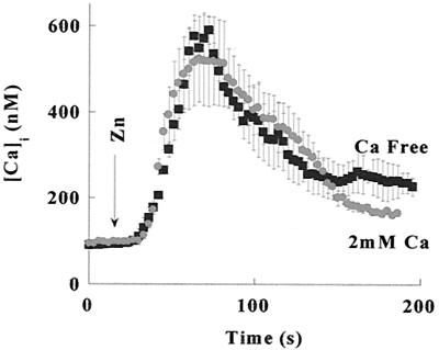
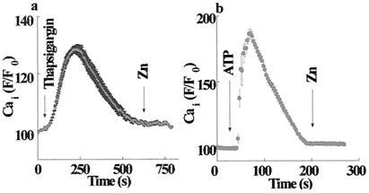
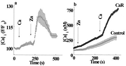
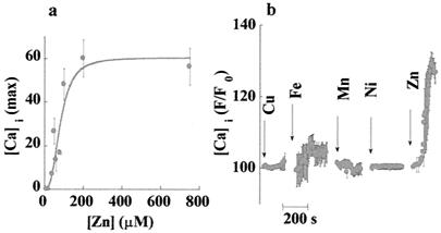
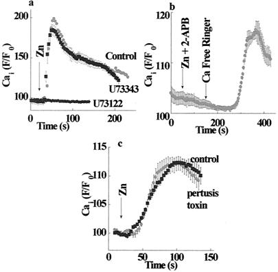
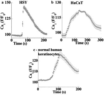
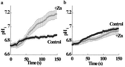
Changes in extracellular zinc concentration participate in modulating fundamental cellular processes such as proliferation, secretion, and ion transport in a mechanism that is not well understood. Here, we show that a micromolar concentration of extracellular zinc triggers a massive release of calcium from thapsigargin-sensitive intracellular pools in the colonocytic cell line HT29. Calcium release was blocked by a phospholipase-C inhibitor, indicating that formation of inositol 1,4,5-triphosphate is required for zinc-dependent calcium release. Zinc influx was not observed, indicating that extracellular zinc triggered the release. The Ca(i)2+ release was zinc specific and could not be triggered by other heavy metals. Furthermore, zinc failed to activate the Ca(2+)-sensing receptor heterologously expressed in HEK293 cells. The zinc-induced Ca(i)2+ rise stimulated the activity of the Na(+)/H(+) exchanger in HT29 cells. Our results indicate that a previously uncharacterized extracellular, G protein-coupled, Zn(2+)-sensing receptor is functional in colonocytes. Because Ca(i)2+ rise is known to regulate key cellular and signal-transduction processes, the zinc-sensing receptor may provide the missing link between extracellular zinc concentration changes and the regulation of cellular processes.


Figures

Figure 1
Figure 1
Cai2+ increase in response to application of zinc. Changes in Cai2+ in HT29 cells loaded with Fura-2 in response to application of Zn2+, as described under Materials and Methods. Zinc (100 μM) was added at the indicated time to HT29 cells superfused with 2 mM Ca2+-containing Ringer's solution (●) or Ca2+-free Ringer's solution (■). In both experiments, application of Zn2+ resulted in a similar increase of Cai2+, indicating that Ca2+ is released from intracellular pools.
Figure 2
Figure 2
Zinc triggers the release of calcium from thapsigargin-sensitive intracellular stores. (a) HT29 cells were superfused with Ca2+-free Ringer's solution and treated with thapsigargin (200 nM), thus inducing a transient Cai2+ rise caused by the emptying of intracellular calcium stores. Zn2+ (200 μM) was added after Cai2+ returned to a resting level. (b) ATP (100 μM) was applied to HT29 cells superfused with Ca2+-free Ringer's solution, resulting in intracellular calcium release; then, Zn2+ (200 μM) was applied (indicated by Zn arrow). No Zn2+-dependent Cai2+ release was observed after the emptying of intracellular stores, thus indicating that extracellular Zn2+ induces the release of Ca2+ from thapsigargin-sensitive intracellular stores.
Figure 3
Figure 3
ZnR is distinct from the CaR. (a) HT29 cells were superfused with 2 mM Ca2+-containing Ringer's solution to activate the CaR. At the time indicated by the arrow, Zn2+ (100 μM) was added to the superfusing solution. The response to Zn2+ was ≈7-fold larger than the response to Ca2+. (b) HEK293 cells transfected with the CaR or the vector alone (Control) were superfused with Ringer's solution containing 0.3 mM Ca2+ and 250 μM Zn2+; then, 2 mM Ca2+ was added to activate the CaR. Cells expressing the CaR did not respond to the application of Zn2+.
Figure 4
Figure 4
Affinity and specificity of the ZnR. (a) Maximal responses to zinc at various concentrations are shown. Each data point represents the means ± SE of five independent experiments. Data were fitted by using the Michaelis–Menten equation, yielding an apparent Km of 80 ± 15 μM. The curve is sigmoidal with a Hill coefficient of 3, which is consistent with the cooperative interaction of zinc ions with ZnR. (b) Cells were superfused with Ca2+-containing Ringer's solution containing 500 μM Mn or Fe; other experiments were done with Ca2+-free Ringer's solution containing 250 μM Cu or Ni (the beginning of each experiment is marked by an arrow). Also, no response was observed when Ni or Cu were introduced in Ca2+-containing Ringer's solution (data not shown). The lack of response to any of the other heavy metals measured indicates that the ZnR was specifically activated by Zn2+.
Figure 5
Figure 5
Signal transduction pathway activated by ZnR. (a) Cells were treated (as marked) with 1 μM PLC inhibitor U73122 (active form), or U73343 (inactive form), or the equivalent volume of DMSO (control), and the calcium response was determined. Zinc-dependent calcium release was inhibited by the active but not by the inactive compound, indicating that ZnR activates the PLC. (b) Cells were pretreated with the IP3 receptor–inhibitor diphenylboric acid 2-aminoethyl ester (2-ApB), and the calcium response was monitored in Ca2+-free Ringer's solution. The zinc-dependent calcium release was blocked in the presence of 2-APB. Washing out the inhibitor with Ca2+-free Ringer's solution resulted in a calcium rise. This fact indicates that ZnR activated the release of IP3, which only was apparent once the inhibition of its receptor was released. The binding of the IP3 to the unblocked receptor resulted in intracellular Ca2+ rise. (c) Controls and cells treated with 50 ng/ml pertussis toxin were superfused with Zn2+ (200 μM). No significant change in ZnR activity was apparent, suggesting that the Go and Gi proteins are not part of the ZnR signal-transduction pathway.
Figure 6
Figure 6
ZnR activity is observed in various cell types. The indicated cells were superfused with calcium-free Ringer's solution followed by the application of 100 μM zinc; changes in Cai2+ were monitored. (a) HSY, human salivary-gland cell line. (b) HaCat, human keratinocytic cell line. (c) For the primary culture of normal human keratinocytes, the graph shows the averaged fluorescence signal of the zinc-responding cells (as described in Results).
Figure 7
Figure 7
Cai2+ rise mediated by ZnR stimulates Na+/H+ exchanger activity. The Na+/H+ exchanger was stimulated by acidifying HT29 cells using an ammonium prepulse protocol. The rate of pHi recovery was determined by monitoring changes in BCECF fluorescence in the presence or absence of 200 μM zinc (as marked). (a) The rate of pHi recovery was accelerated ≈5-fold in the presence of Zn2+. The rate of pHi recovery was 15.7 ± 0.1 × 10⁻³ pHi units per s in the presence of Zn2+, as compared with 3.0 ± 0.1 × 10⁻³ pHi units per s in the control. (b) Cells treated with thapsigargin (200 nM) were superfused with calcium-free Ringer's solution. The rate of pHi recovery after thapsigargin treatment was 3.5 ± 0.4 × 10⁻³ pHi units per s in the presence of Zn2+, as compared with 2.7 ± 0.4 × 10⁻³ pHi units per s in the control. Eliminating the changes in Cai2+ inhibited the zinc-stimulated pHi recovery. These results indicate that the stimulation of the Na+/H+ exchanger by the ZnR is mediated by the zinc-induced rise in Cai2+.

Comment in

References

    1. Berg J M, Shi Y. Science. 1996;271:1081–1085. - PubMed
    1. Vallee B L, Falchuk K H. Physiol Rev. 1993;73:79–118. - PubMed
    1. Huang E P. Proc Natl Acad Sci USA. 1997;94:13386–13387. - PMC - PubMed
    1. Wapnir R A. J Nutr. 2000;130:1388S–1392S. - PubMed
    1. Prasad A S. Nutrition. 1995;11:93–99. - PubMed

Publication types

MeSH terms

Substances

LinkOut - more resources