. 2001 Sep 25;98(20):11806-11.
doi: 10.1073/pnas.201413498.

LEAFY COTYLEDON2 encodes a B3 domain transcription factor that induces embryo development

Affiliations

LEAFY COTYLEDON2 encodes a B3 domain transcription factor that induces embryo development

S L Stone et al. Proc Natl Acad Sci U S A. .

Abstract

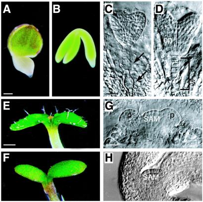
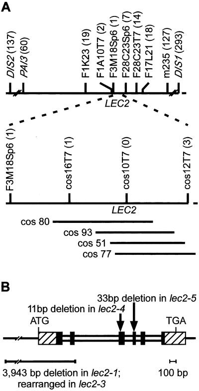
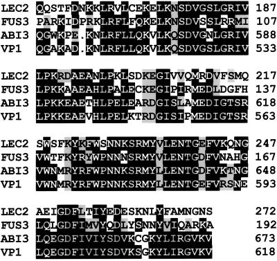
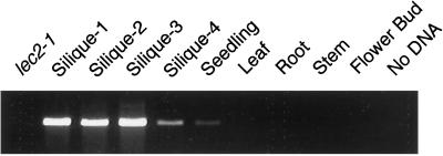
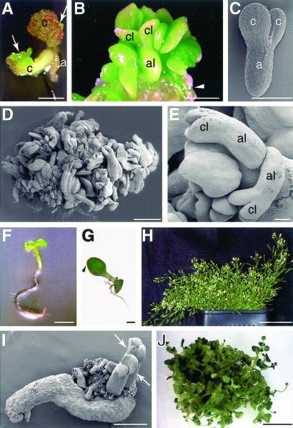
The Arabidopsis LEAFY COTYLEDON2 (LEC2) gene is a central embryonic regulator that serves critical roles both early and late during embryo development. LEC2 is required for the maintenance of suspensor morphology, specification of cotyledon identity, progression through the maturation phase, and suppression of premature germination. We cloned the LEC2 gene on the basis of its chromosomal position and showed that the predicted polypeptide contains a B3 domain, a DNA-binding motif unique to plants that is characteristic of several transcription factors. We showed that LEC2 RNA accumulates primarily during seed development, consistent with our finding that LEC2 shares greatest similarity with the B3 domain transcription factors that act primarily in developing seeds, VIVIPAROUS1/ABA INSENSITIVE3 and FUSCA3. Ectopic, postembryonic expression of LEC2 in transgenic plants induces the formation of somatic embryos and other organ-like structures and often confers embryonic characteristics to seedlings. Together, these results suggest that LEC2 is a transcriptional regulator that establishes a cellular environment sufficient to initiate embryo development.


Figures

Figure 1
Figure 1
Morphological phenotype of lec2 mutants. (A) lec2-5, (C) lec2-3, and (E, G) lec2-4 mutant embryos. (B, D, F, H) Wild-type embryo. (A and B) Whole-mount photographs of maturing embryos. (C and D) Embryonic suspensors as viewed by using differential interference contrast (DIC) microscopy of cleared seeds. Arrows point to abnormal suspensor cells in lec2 mutants. (E and F) Cotyledons of seedlings grown for 4–5 days. A lec2 mutant seedling germinated before desiccation possessed trichomes on the adaxial surface of cotyledons. (G and H) Shoot apices of curled cotyledon-stage embryos seen with DIC optics. The shoot apical meristem of lec2 mutants is domed and possesses leaf primordia in contrast to the unactivated meristem of wild types. ep, Embryo proper; p, leaf primordium; SAM, shoot apical meristem; s, suspensor. [Bars = 100 μm (A), 20 μm (C, G), 300 μm (E).]
Figure 2
Figure 2
Genetic mapping and positional cloning of the LEC2 gene. (A) Diagrammatic representation of the interval on chromosome 1 between DIS1 and DIS2. The position of the LEC2 gene relative to genetic markers, including the ends of BAC and cosmid clones, are indicated. Numbers in parentheses show recombinant breakpoints observed between the indicated marker and LEC2. Positions of cosmid clones that suppress the lec2 mutation are shown. (B) Representation of the LEC2 gene. Hatched and shaded boxes indicate exons, and narrow boxes represent introns. Shaded boxes depict the gene region encoding the B3 domain. The position of mutations in specific mutant alleles are indicated. Rearrangement of lec2-3 was assessed with DNA gel blot hybridization studies.
Figure 3
Figure 3
LEC2 contains a B3 domain. Amino acid alignment of residues from the B3 domains of LEC2, FUS3, ABI3, and VP1. Residues in black boxes are identical in at least two of the four proteins, and those in shaded boxes share similarity with conserved residues. Numbers in the right column indicate residue numbers in the predicted polypeptides.
Figure 4
Figure 4
LEC2 RNA accumulates primarily during seed development. LEC2 RNA was detected at the indicated stages by amplifying reverse transcription products. Silique stages 1 through 4, respectively, are from siliques containing zygote to early globular-stage embryos, globular-stage to heart-stage embryos, torpedo-stage to curled cotyledon-stage embryos, and mature green embryos. Control experiments showed that a ribosomal protein RNA was amplified with similar efficiency from each reverse transcription reaction.
Figure 5
Figure 5
LEC2 induces somatic embryo development. Seeds from lec2-1 and lec2-5 plants transformed with the 35SLEC2 gene were germinated, and their subsequent development was monitored. (A) Formation of somatic embryo-like clusters (arrows) on the cotyledons of an embryo-like 35SLEC2 seedling. (B) Somatic embryo-like structures emerging from the cotyledon (arrowhead) of a 35SLEC2 seedling. (C) Scanning electron microscopy (SEM) photograph of a wild-type linear cotyledon-stage zygotic embryo. (D) A mass of somatic embryo-like structures covering a 35SLEC2 seedling as observed by SEM. (E) SEM photograph of somatic embryo-like structures on a 35SLEC2 seedling. (F) Seedling resulting from the germination of a 35SLEC2 somatic embryo formed on a wild-type transgenic seedling. (G) A 35SLEC2 seedling with a wild-type phenotype. Arrowhead shows that the distal region of the cotyledon is not defective. (H) A short, bushy plant grown on soil that developed from a 35SLEC2 seedling such as that shown in G. (I) Somatic embryos (arrows) emerging from leaf-like organ dissected from a plantlet mass. (J) A mass of plantlets formed from a single 35SLEC2 seedling such as that shown in G. a, Embryonic axis; al, embryonic axis-like; c, cotyledon; cl, cotyledon-like. [Bars = 1 mm (A, B, D, F, G), 100 μm (C, E), 500 μm (I), 1 cm (J), and 5 cm (H).]
Figure 6
Figure 6
LEC2-induced somatic embryos express embryo-specific genes. (A) Whole-mount photograph of a 35SLEC2 seedling with somatic embryo-like structures on its cotyledons. Sections of the same seedling were hybridized with antisense probes for LEC2 RNA (B), cruciferin A storage protein RNA (C), and oleosin lipid body protein RNA (D). After autoradiography, sections were photographed with use of dark-field optics. A sense-strand probe did not hybridize appreciably with the sections (data not shown). Regions of the seedlings in B–D that did not hybridize lacked cellular contents and were likely dead. c, Cotyledon; se, somatic embryo. (Bars = 0.5 mm.)

Similar articles

Cited by

References

    1. Olsen O A. Annu Rev Plant Physiol Plant Mol Biol. 2001;52:241–267. - PubMed
    1. Goldberg R B, de Paiva G, Yadegari R. Science. 1994;266:605–614. - PubMed
    1. West M A, Harada J J. Plant Cell. 1993;5:1361–1369. - PMC - PubMed
    1. Jurgens G. EMBO J. 2001;20:3609–3616. - PMC - PubMed
    1. Bewley J D. Plant Cell. 1997;9:1055–1066. - PMC - PubMed

MeSH terms

Associated data