. 2001 Oct;8(10):874-8.
doi: 10.1038/nsb1001-874.

Interaction of coxsackievirus B3 with the full length coxsackievirus-adenovirus receptor

Affiliations

Interaction of coxsackievirus B3 with the full length coxsackievirus-adenovirus receptor

Y He et al. Nat Struct Biol. 2001 Oct.

Abstract

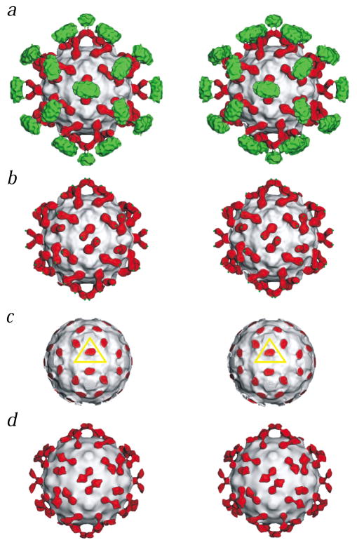
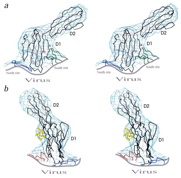
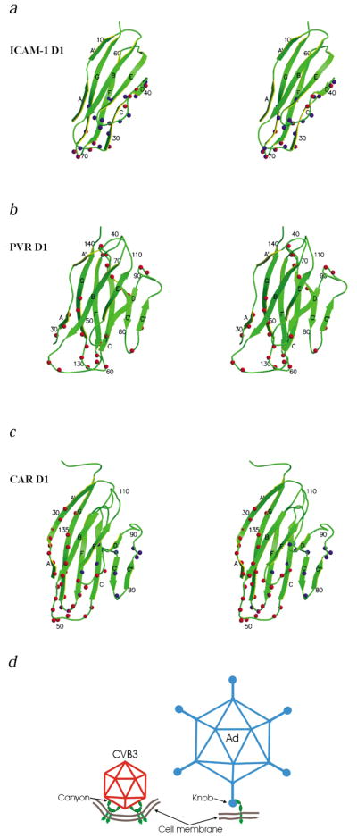
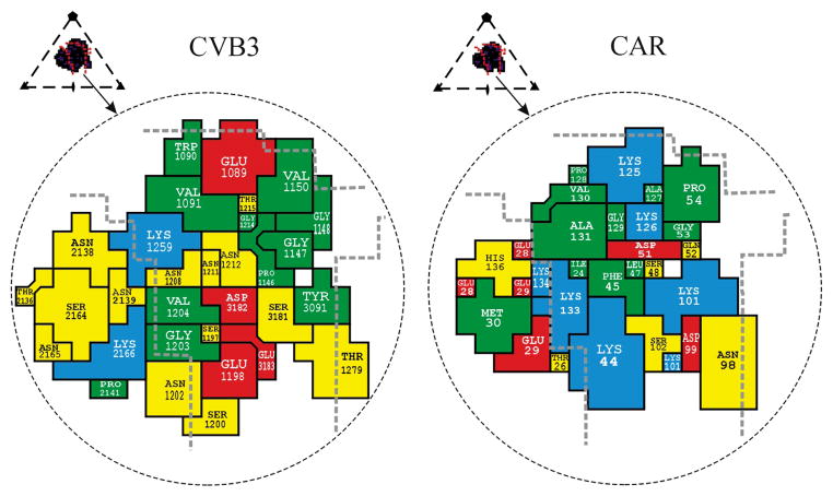
Group B coxsackieviruses (CVB) utilize the coxsackievirus-adenovirus receptor (CAR) to recognize host cells. CAR is a membrane protein with two Ig-like extracellular domains (D1 and D2), a transmembrane domain and a cytoplasmic domain. The three-dimensional structure of coxsackievirus B3 (CVB3) in complex with full length human CAR and also with the D1D2 fragment of CAR were determined to approximately 22 A resolution using cryo-electron microscopy (cryo-EM). Pairs of transmembrane domains of CAR associate with each other in a detergent cloud that mimics a cellular plasma membrane. This is the first view of a virus-receptor interaction at this resolution that includes the transmembrane and cytoplasmic portion of the receptor. CAR binds with the distal end of domain D1 in the canyon of CVB3, similar to how other receptor molecules bind to entero- and rhinoviruses. The previously described interface of CAR with the adenovirus knob protein utilizes a side surface of D1.


Figures

Fig. 1
Fig. 1
Stereo views of CAR bound to CVB3. The virus in each panel is represented as a grayscale surface. Domains D1 and D2 of CAR are colored red, and the transmembrane and cytoplasmic regions are green. a, CVB3 complexed with the full length CAR. b, Same as (a) but with density outside a radius of 200 Å removed to show domains D1 and D2 of CAR only (green dots indicate where the transmembrane regions start). c, Same as (a) but with density removed outside a radius of 154 Å to show the binding site of D1 in the CVB3 canyon. The yellow triangle defines the icosahedral asymmetric unit. d, The complex of CVB3 with the CAR D1D2 fragment.
Fig. 2
Fig. 2
Orthogonal stereo views of the Cα backbone of CAR D1 and D2 (black) fit into the cryo-EM density. Fragments of CVB3 (blue for VP1, green for VP2 and red for VP3) are also shown. a, The south rim of the canyon, formed by VP2, contacts the A and G β-strands of domain D1. b, The sugar moieties of the carbohydrate at Asn 108 are depicted in yellow.
Fig. 3
Fig. 3
Stereo diagrams of the ICAM-1, PVR and CAR D1 domains. The β-strands are labeled A–G. The amino acids identified as being in the virus–receptor interface are indicated by spheres. a, ICAM-1 with HRV14 and HRV16 in blue, and ICAM-1 with coxsackievirus A21 in red; b, PVR with poliovirus in red; and c, CAR with adenovirus knob in blue and CAR with CVB3 in red. d, Schematic diagram of the modes by which CAR (green) binds to CVB3 (red) and adenovirus (blue). The suggested membrane curvature is speculative.
Fig. 4
Fig. 4
Footprint of domain D1 onto the CVB3 surface (left) and footprint of CVB3 onto CAR (right). Inset shows the icosahedral asymmetric unit of the virus, the rough boundaries of the canyon (red lines) and the perimeter of the receptor footprint. The amino acids forming the interface are colored as follows: red for acidic residues, blue for basic residues, yellow for polar residues and green for hydrophobic residues. Thick dashed gray lines highlight the canyon boundaries. The CAR amino acids are numbered in accordance with the system used by Bewley et al. (PDB 1KAC); these sequence numbers are greater by two than those used by van Raaij et al. (PDB 1F5W).

References

    1. Rueckert RR. In: Fields virology. Fields BN, Knipe DM, Howley PM, editors. Lippincott-Raven Press; Philadelphia & New York: 1996. pp. 609–654.
    1. Melnick JL. In: Fields virology. Fields BN, Knipe DM, Howley PM, editors. Lippincott-Raven Publishers; Philadelphia & New York: 1996. pp. 655–712.
    1. Muckelbauer JK, et al. Structure. 1995;3:653–668. - PubMed
    1. Lonberg-Holm K, Crowell RL, Philipson L. Nature. 1976;259:679–681. - PubMed
    1. Bergelson JM, et al. Science. 1997;275:1320–1323. - PubMed

Publication types

Substances

Associated data

LinkOut - more resources