. 2001 Oct 1;20(19):5332-41.
doi: 10.1093/emboj/20.19.5332.

IL-17s adopt a cystine knot fold: structure and activity of a novel cytokine, IL-17F, and implications for receptor binding

Affiliations

IL-17s adopt a cystine knot fold: structure and activity of a novel cytokine, IL-17F, and implications for receptor binding

S G Hymowitz et al. EMBO J. .

Abstract

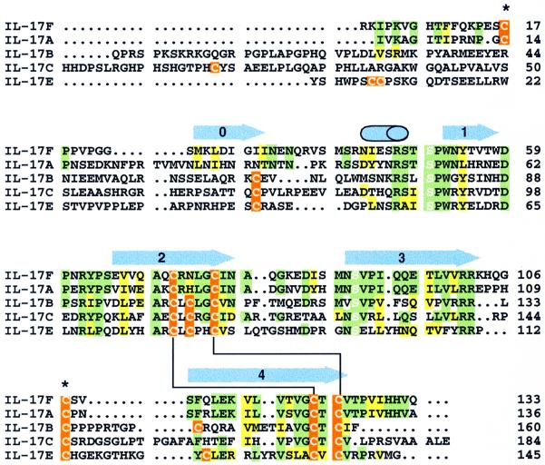
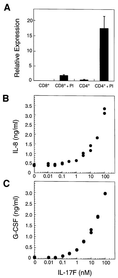
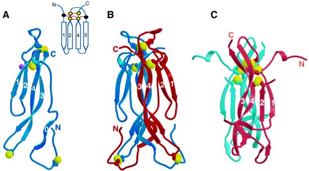
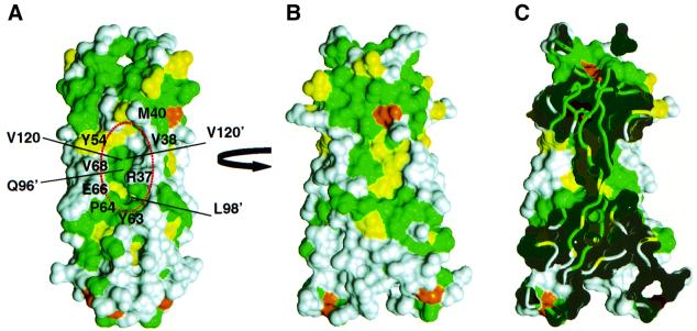
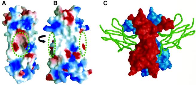
The proinflammatory cytokine interleukin 17 (IL-17) is the founding member of a family of secreted proteins that elicit potent cellular responses. We report a novel human IL-17 homolog, IL-17F, and show that it is expressed by activated T cells, can stimulate production of other cytokines such as IL-6, IL-8 and granulocyte colony-stimulating factor, and can regulate cartilage matrix turnover. Unexpectedly, the crystal structure of IL-17F reveals that IL-17 family members adopt a monomer fold typical of cystine knot growth factors, despite lacking the disulfide responsible for defining the canonical "knot" structure. IL-17F dimerizes in a parallel manner like neurotrophins, and features an unusually large cavity on its surface. Remarkably, this cavity is located in precisely the same position where nerve growth factor binds its high affinity receptor, TrkA, suggesting further parallels between IL-17s and neurotrophins with respect to receptor recognition.


Figures

None
Fig. 1. Sequence alignment of IL-17F with other IL-17 family members. Regions of sequence identity or similarity between IL-17F and IL-17A are highlighted in green and yellow, respectively. When the other family members also have an identical or similar residue at the same position, they are similarly colored. Cysteine residues are indicated in orange. The conserved serines that replace the canonical knot cysteines are highlighted with white letters. Disulfide bonds that are expected to be conserved in all IL-17s are indicated by a black line connecting the bonded cystines. The two cysteines that form the inter-chain disulfide in IL-17F are marked with an asterisk. Secondary structural elements in IL-17F are shown above the sequences as blue arrows (β-strands) or a cylinder (α-helix). Residue numbering is from the start of the mature sequences.
None
Fig. 2. IL-17F is produced by activated T cells and stimulates cytokine production. (A) IL-17F expression in T cells. Relative mRNA expression is shown. PI, treated with PMA and ionomycin. Induction of IL-8 (B) and G-CSF (C) in fibroblasts by IL-17F. Human primary foreskin fibroblasts were cultured for 24 h in the presence of the indicated concentrations of IL-17F. Conditioned medium was then analyzed by ELISA for the presence of IL-8 and G-CSF.
None
Fig. 3. Effect of IL-17F on porcine and human cartilage. Porcine articular cartilage (AC) and human articular cartilage (from 65-year-old caucasian female) (DF) explants were treated with varying concentrations (0.1, 1 or 10 nM) of IL-17F or IL-17A, or 0.1 nM human IL-1α, and proteoglycan breakdown (A and D), proteoglycan synthesis (B and E) and IL-6 production (C and F) were measured. Data represent the average of five independent samples ± SEM. ‘0’ in each panel represents the untreated control. *denotes statistically significant difference from control (p<0.05).
None
Fig. 4. The structure of IL-17F. (A) Ribbon trace of the IL-17F monomer. Strands are labeled. Disulfides are shown as ball-and-stick representation with the sulfur atoms colored yellow. Glycosylation of Asn53 is indicated by a purple ball. Inset shows a cartoon representation of the canonical cystine knot fold. Cysteine residues are indicated by filled circles; those present in IL-17 proteins are yellow, whereas the two that are missing are black. (B) Ribbon trace of the IL-17F dimer. Disulfides are shown as in (A). (C) The structure of NGF from the NGF–TrkA complex (Wiesmann et al., 1999; Protein Data Bank code 1WWW); a disordered loop connects strands 2 and 3.
None
Fig. 5. Comparison of IL-17F and IL-17A. Two orthogonal views, ‘side’ (A) and ‘front’ (B), of the molecular surface of IL-17F colored according to sequence conservation between IL-17A and F as in Figure 1. The surface of residues that are identical between the two proteins are colored green, homologous residues are colored yellow, while residues that differ significantly are colored white. [The view in (B) is oriented ∼15° rotated from the view in Figure 4B.] Residues forming the cavity are labeled in (A). (C) ‘Cut-away’ view of the surface in (B) showing how the large cavities on either side of IL-17F penetrate deeply into the body of the dimer.
None
Fig. 6. Comparison of the IL-17F surface and the TrkA-binding site on NGF. (A and B) The molecular surface of IL-17F is oriented as in Figure 5. IL-17F is colored according to the electrostatic surface potential: red, –5 kT; white, 0 kT; and blue, +5 kT. The positions of the cavities are indicated by the circles. (C) The molecular surface of NGF is shown in the same orientation as IL-17F in (B) with the two protomers of the dimer colored red and blue; domain 5 of TrkA is shown as a green ribbon (Wiesmann et al., 1999).

References

    1. Arend W.P. and Dayer,J.-M. (1990) Cytokines and cytokine inhibitors or antagonists in rheumatoid arthritis. Arthritis Rheum., 33, 305–315. - PubMed
    1. Awane M., Andres,P.G., Li,D.J. and Reinecker,H.-C. (1999) NF-κB-inducing kinase is a common mediator of IL-17-, TNF-α-, and IL-1β-induced chemokine promoter activation in intestinal epithelial cells. J. Immunol., 162, 5337–5344. - PubMed
    1. Banner D.W., D’Arcy,A., Janes,W., Gentz,R., Schoenfeld,H.-J., Broger,C., Loetscher,H. and Lesslauer,W. (1993) Crystal structure of the soluble human 55 kd TNF receptor–human TNFβ complex: implications for TNF receptor activation. Cell, 73, 431–445. - PubMed
    1. Bowie A. and O’Neill,L.A.J. (2000) The interleukin-1 receptor/Toll-like receptor superfamily: signal generators for pro-inflammatory interleukins and microbial products. J. Leukoc. Biol., 67, 508–514. - PubMed
    1. Brünger A.T. (1992) X-PLOR Manual, Version 3.1. Yale University Press, New Haven, CT.

MeSH terms

Associated data