. 2001 Oct 1;20(19):5347-53.
doi: 10.1093/emboj/20.19.5347.

NMR structure of the LCCL domain and implications for DFNA9 deafness disorder

Affiliations

NMR structure of the LCCL domain and implications for DFNA9 deafness disorder

E Liepinsh et al. EMBO J. .

Abstract

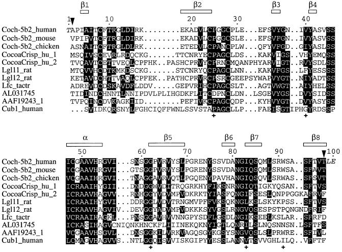
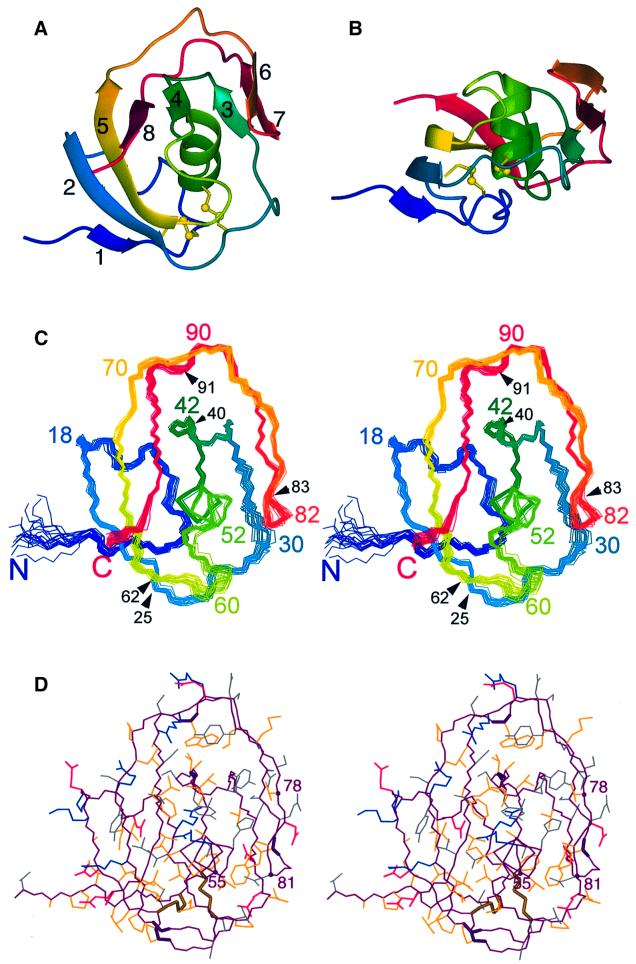
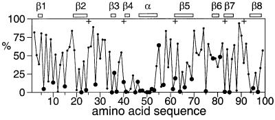
The LCCL domain is a recently discovered, conserved protein module named after its presence in Limulus factor C, cochlear protein Coch-5b2 and late gestation lung protein Lgl1. The LCCL domain plays a key role in the autosomal dominant human deafness disorder DFNA9. Here we report the nuclear magnetic resonance (NMR) structure of the LCCL domain from human Coch-5b2, where dominant mutations leading to DFNA9 deafness disorder have been identified. The fold is novel. Four of the five known DFNA9 mutations are shown to involve at least partially solvent-exposed residues. Except for the Trp91Arg mutant, expression of these four LCCL mutants resulted in misfolded proteins. These results suggest that Trp91 participates in the interaction with a binding partner. The unexpected sensitivity of the fold with respect to mutations of solvent-accessible residues might be attributed to interference with the folding pathway of this disulfide-containing domain.


Figures

None
Fig. 1. Multiple alignment of the amino acid sequences of LCCL domains. The top sequence is the construct used in the present study. The residues of the construct are numbered from 1 to 100. This numbering is used throughout this article and differs by 26 from the residue numbering by Trexler et al. (2000). The construct thus comprises residues 28–124 of human Coch-5b2 (positions indicated by arrows) and contains three additional residues at the termini from the expression system used (shown in italics). The locations of the α-helix and β-strands in human Coch-5b2 are shown at the top. The following sequences are from mouse Coch-5b2 (Coch-5b2_mouse, residues 30–126), chicken Coch-5b2 (Coch-5b2_chicken, residues 24–120), the two LCCL domains of the human CocoaCrisp protein (CocoaCrisp_hu_1, residues 289–387, and CocoaCrisp_hu_2, residues 390–497), the two LCCL domains of rat late gestation lung protein Lgl1 (Lgl11_rat, residues 224–322, and Lgl12_rat, residues 325–433), Limulus factor C (Lfc_tactr, residues 325–424), a predicted protein of P. falciparum (DDBJ/EMBL/GenBank accession No. AL031745), the predicted human Coch-5b2-related protein (DDBJ/EMBL/GenBank accession No. AAF19243, residues 40–136) and the predicted human Cub-1 protein (Cub1_human, residues 116–213). To highlight conserved features of the LCCL domain, similar residues present in more than half of the LCCL modules are shaded. Crosses mark the locations of residues, where mutations in the human Coch-5b2 protein correlate with the deafness disorder DFNA9.
None
Fig. 2. Solution structure of the LCCL domain of human Coch-5b2. (A) Ribbon representation. The disulfide bonds are shown in a ball-and-stick representation. The β-strands are numbered as in Figure 1. (B) Ribbon representation as in (A), but in a different orientation. (C) Stereo view showing a superposition of the backbone atoms in the 20 conformers representing the NMR structure (Table I), in a similar orientation to that in (A). Approximately every tenth residue is identified by its sequence number. In addition, black arrows and sequence numbers identify the locations of the five known DFNA9 mutations. (D) Stereo view of the conformer closest to the mean structure of the 20 conformers shown in (C), using a heavy atom representation in the same orientation as in (C). The polypeptide backbone is drawn in purple. The following colors were used for the side chains: blue, Arg, Lys; red, Glu, Asp; yellow, Ala, Cys, Ile, Leu, Met, Phe, Pro, Trp, Val; gray, Asn, Gln, His, Ser, Thr, Tyr. Bold lines label the backbone of the residues mutated in the deafness disorder DFNA9 (Pro25, Val40, Gly62, Ile 83, Trp91) and the two disulfide bridges. Spheres and residue numbers highlight the Cα atoms of the fully conserved residues Gly55 and Asn81, and of residue 78, where all LCCL domains have an uncharged and solvent-exposed residue (Figures 1 and 3).
None
Fig. 3. Solvent accessibility of the amino acid side chains in the LCCL domain of human Coch-5b2. The solvent accessibilities are expressed as a percentage of their accessibilities calculated for the situation where the respective amino acid residues are located in a hypothetical poly-Gly α-helix with a fully extended side chain (Sevilla-Sierra et al., 1994). The values were averaged over the 20 NMR conformers. Large filled circles identify highly conserved amino acid residues that are uncharged in all LCCL domains known to date (Figure 1), including His in the group of uncharged residues. The locations of the α-helix and β-strands are indicated at the top. Crosses identify the locations of DFNA9 mutations.

References

    1. Andersson P., Weigelt,J. and Otting,G. (1998) Spin-state selection filters for the measurement of heteronuclear one-bond coupling constants. J. Biomol. NMR, 12, 435–441. - PubMed
    1. Bartels C., Xia,T., Güntert,P., Billeter,M. and Wüthrich,K. (1995) The program XEASY for computer-supported NMR spectral analysis. J. Biomol. NMR, 5, 1–10. - PubMed
    1. Bom S.J., Kemperman,M.H., de Kok,Y.J., Huygen,P.L., Verhagen,W.I., Cremers,F.P. and Cremers,C.W. (1999) Progressive cochleo-vestibular impairment caused by a point mutation in the COCH gene at DFNA9. Laryngoscope, 109, 1525–1530. - PubMed
    1. Colombatti A. and Paolo,B. (1991) The superfamily of proteins with von Willebrand factor type A-like domains: one theme common to components of extracellular matrix, hemostasis, cellular adhesion, and defense mechanisms. Blood, 77, 2305–2315. - PubMed
    1. Colombatti A., Bonaldo,P. and Doliana,R. (1993) Type A modules: interacting domains found in several non-fibrillar collagens and in other extracellular matrix proteins. Matrix, 13, 297–306. - PubMed

Publication types

Associated data