. 2001 Oct 1;20(19):5400-11.
doi: 10.1093/emboj/20.19.5400.

Direct interaction between the Arabidopsis disease resistance signaling proteins, EDS1 and PAD4

Affiliations

Direct interaction between the Arabidopsis disease resistance signaling proteins, EDS1 and PAD4

B J Feys et al. EMBO J. .

Abstract

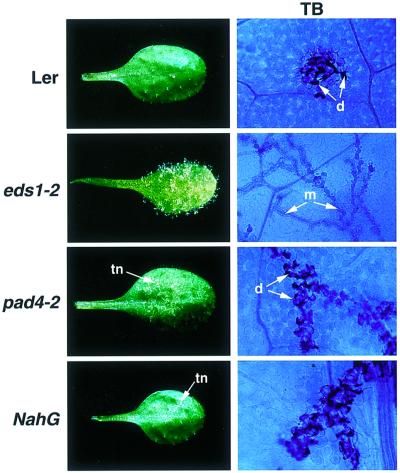
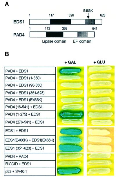
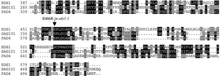
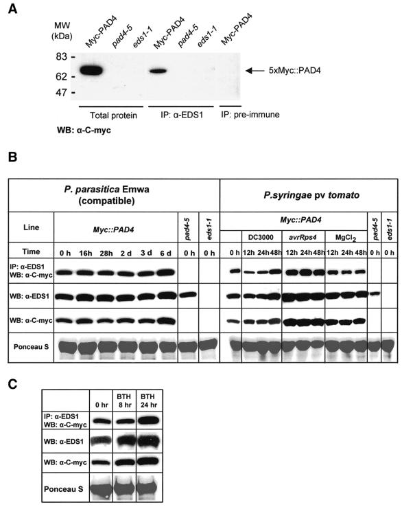
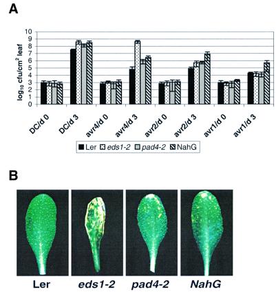
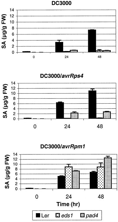
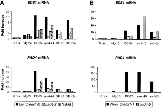
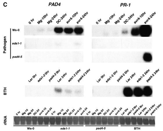
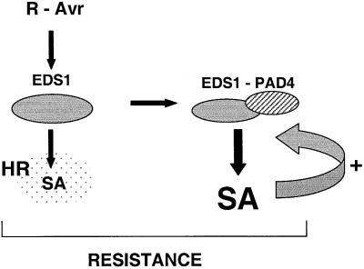
The Arabidopsis EDS1 and PAD4 genes encode lipase-like proteins that function in resistance (R) gene-mediated and basal plant disease resistance. Phenotypic analysis of eds1 and pad4 null mutants shows that EDS1 and PAD4 are required for resistance conditioned by the same spectrum of R genes but fulfil distinct roles within the defence pathway. EDS1 is essential for elaboration of the plant hypersensitive response, whereas EDS1 and PAD4 are both required for accumulation of the plant defence-potentiating molecule, salicylic acid. EDS1 is necessary for pathogen-induced PAD4 mRNA accumulation, whereas mutations in PAD4 or depletion of salicylic acid only partially compromise EDS1 expression. Yeast two-hybrid analysis reveals that EDS1 can dimerize and interact with PAD4. However, EDS1 dimerization is mediated by different domains to those involved in EDS1-PAD4 association. Co-immunoprecipitation experiments show that EDS1 and PAD4 proteins interact in healthy and pathogen-challenged plant cells. We propose two functions for EDS1. The first is required early in plant defence, independently of PAD4. The second recruits PAD4 in the amplification of defences, possibly by direct EDS1-PAD4 association.


Figures

None
Fig. 1. RPP5 resistance phenotypes of wild-type Ler, eds1-2, pad4-2 and NahG leaves inoculated with P.parasitica isolate Noco2. Two-week-old seedlings were spray-inoculated with a suspension of P.parasitica conidia (5 × 10⁴/ml) and incubated as described in Materials and methods. Whole leaves were photographed 6 days after inoculation. Trailing necrosis (tn) in pad4-2 and NahG is indicated by an arrow. Leaf tissue was stained with lactophenol Trypan Blue (TB) 5 days after inoculation to visualize pathogen mycelium (m) and necrotic plant cells (d). The TB-stained material, viewed under a light microscope, is shown at ×400 magnification.
None
Fig. 2. Interaction between EDS1 and PAD4 in a yeast two-hybrid assay. (A) Schematic representation of the domain structure of the Arabidopsis EDS1 and PAD4 proteins. The lipase domain (filled box) and EP (EDS1 and PAD4-defined) domain (hatched box) are indicated. The position of the eds1-1 (E466K) mutation is shown with an arrow. The EP domain lies between residues 405 and 554 (EDS1) and residues 332 and 457 (PAD4). (B) Two-hybrid interactions between EDS1 and PAD4. Full-length proteins or defined subdomains of EDS1 and PAD4 were tested for specific interactions under inducing (+GAL) or repressing conditions (+GLU). Combinations are shown with the first protein fused to the LexA domain and the second partner fused to the AD domain. Numbers refer to amino acid positions in the full-length protein. Positive interactions are defined by activation of the LacZ (shown) and LEU2 (same pattern as LacZ; data not shown) reporter genes. The interaction between p53 and SV40-T serves as a positive control for the assay.
None
Fig. 3. Sequence alignment of the EP domain in EDS1, PAD4 and SAG101. Sequences of the Arabidopsis EDS1 and PAD4 proteins were aligned with SAG101, a senescence-associated gene of unknown function (He et al., 2001) across the EP domain. The position of the eds1-1 (E466K) mutation is indicated with an arrow. The alignment was generated using Clustal_W and shaded using BoxShade (see Materials and methods). Positions are relative to the full-length protein. The DDBJ/EMBL/GenBank accession No. for SAG101 is AAF78583.
None
Fig. 4. In planta protein interaction between EDS1 and PAD4. (A) Co-immunoprecipitation of EDS1 and PAD4 in total plant protein extracts. Protein extracts were prepared from the transgenic pad4-5 (5× Myc::PAD4) line, indicated as Myc-PAD4, or from the pad4-5 and eds1-1 mutants. For immunoprecipitation reactions pre-immune (as control) or EDS1 antiserum was used, followed by western blotting detection with anti-c-Myc antibody. Total protein extracts were analyzed on the same western blot to show the specificity of the anti-c-Myc antibody. (B) Analysis of EDS1 and PAD4 protein expression and co-immunoprecipitation in healthy and pathogen-challenged plants. Leaves of the 5× Myc::PAD4 epitope-tagged transgenic line were spray inoculated with P.parasitica spores (1 × 10⁵/ml in dH20) or infiltrated with suspensions (5 × 10⁶/ml colony forming units in 10 mM MgCl2) of DC3000, DC3000 expressing avrRps4 or 10 mM MgCl2 alone, and tissues harvested at the time points indicated. Levels of EDS1 and PAD4 protein were measured on western blots of total soluble extracts probed with anti-EDS1 and anti-c-Myc antibody, respectively. Co-immunoprecipitations were performed on the same tissue extracts, as described in (A). Equal loading of blots is indicated by Ponceau S staining of total protein. An independent experiment gave similar results. (C) Analysis of EDS1 and PAD4 protein expression and co-immunoprecipitation in leaves treated with BTH. Tissues were harvested and analyzed as described in (B). Similar results were obtained in an independent experiment.
None
Fig. 5. Growth and symptom development of different P.syringae strains in leaves of wild-type Ler, eds1-2, pad4-2 and NahG plants. (A) Leaves of 4-week-old short day grown plants were infiltrated with a suspension (1 × 10⁵ colony forming units/ml) of P.syringae pv. tomato strain DC3000 containing an empty vector (DC3000) or DC3000 expressing avrRps4, avrRpt2 or avrRpm1. Bacterial titres were measured at 0 and 3 days after inoculation. The measurements and standard errors are derived from four replicates per treatment. An independent experiment gave similar results. (B) Leaves were dipped in a suspension (1 × 10⁷ c.f.u./ml) of DC3000 expressing avrRps4 and disease symptoms observed over 6 days. As shown at day 5, Ler plants appear healthy, eds1-2 plants develop severe leaf spotting symptoms, while pad4-2 and NahG plants exhibit mild leaf spotting.
None
Fig. 6. Accumulation of total salicylic acid in Ler, eds1-2 and pad4-2 plants after inoculation with virulent and avirulent P.syringae pv. tomato DC3000 strains Leaves of 4-week-old short day grown plants were dipped in a suspension (1 × 10⁷ c.f.u./ml) of DC3000 (top panel), DC3000 expressing avrRps4 (middle panel) or avrRpm1 (bottom panel). Total salicylic acid (SA) was extracted and quantified after 0, 24 and 48 h by HPLC as described in Materials and methods. Salicylic acid measurements and standard errors are derived from three replicate samples per treatment. Salicylic acid was present in trace amounts in Ler-NahG plants at all stages of infection (data not shown).
None
Fig. 7. Abundance of EDS1, PAD4 and PR-1 mRNAs in pathogen-inoculated and BTH-treated plants. (A) Leaves of 5-week-old Ler, eds1-2, pad4-2 and Ler-NahG plants were hand-infiltrated with 10 mM MgCl2 (Mg), 1 × 10⁷ c.f.u./ml P.syringae DC3000 (DC) or P.syringae DC3000 expressing avrRps4 (avr4), or sprayed with 300 µM benzothiadiazole (BTH). Material was harvested after 24 h for the bacterial inoculations and 6 and 24 h for the BTH treatment. Messenger RNA abundance was determined using TaqMan chemistry (see Materials and methods). EDS1 and PAD4 mRNA levels are normalized relative to the internal control ACT2, and are calculated relative to expression at 0 h. (B) Leaves of 5-week-old Ws-0, eds1-1 and pad4-5 plants were pathogen challenged as in (A). Material was harvested 10 and 24 h after challenge. PAD4 mRNA is undetectable in the pad4-5 mutant. Relative quantification using TaqMan chemistry is as described in (A). (C) RNA gel blot analysis of PAD4 and PR-1 mRNA expression. Samples from (B) plus BTH-treated samples from (A) were analysed to verify TaqMan results for PAD4 and examine the expression of PR-1. Results for PAD4 (left) and PR-1 (right) are shown after pathogen challenge (top panel) and BTH treatment (middle panel). Control for equal loading is shown in the bottom panel (rRNA).
None
Fig. 8. A model for the roles of EDS1 and PAD4 in R gene-mediated resistance. Two functions are proposed for EDS1 in R-Avr protein-triggered resistance at pathogen infection foci. One lies upstream of the plant HR (indicated by the stipled area) and is required for a low level of SA accumulation. The second function recruits PAD4, possibly through direct EDS1–PAD4 interaction, and drives amplification of local defences through enhanced accumulation of SA and other molecules (indicated by the curved arrow). Complete containment of the pathogen requires both the HR and defence signal potentiation.

References

    1. Aarts N., Metz,M., Holub,E., Staskawicz,B.J., Daniels,M.J. and Parker,J.E. (1998) Different requirements for EDS1 and NDR1 by disease resistance genes define at least two R gene-mediated signaling pathways in Arabidopsis. Proc. Natl Acad. Sci. USA, 95, 10306–10311. - PMC - PubMed
    1. An Y.Q., McDowell,J.M., Huang,S.R., McKinney,E.C., Chambliss,S. and Meagher,R.B. (1996) Strong, constitutive expression of the Arabidopsis ACT2/ACT8 actin subclass in vegetative tissues. Plant J., 10, 107–121. - PubMed
    1. Ausubel F.M., Brent,R., Kingston,R.E., Moore,D.D., Seidman,J.G., Smith,J.A. and Struhl,K. (1996) Current Protocols in Molecular Biology. John Wiley & Sons, New York, NY.
    1. Bergö M., Olivecrona,G. and Olivecrona,T. (1996) Forms of lipoprotein lipase in rat tissues: in adipose tissue the proportion of inactive lipase increases on fasting. Biochem. J., 313, 893–898. - PMC - PubMed
    1. Botella M.A., Parker,J.E., Frost,L.N., BittnerEddy,P.D., Beynon,J.L., Daniels,M.J., Holub,E.B. and Jones,J.D.G. (1998) Three genes of the Arabidopsis RPP1 complex resistance locus recognize distinct Peronospora parasitica avirulence determinants. Plant Cell, 10, 1847–1860. - PMC - PubMed

Publication types

MeSH terms