. 2001 Oct 1;20(19):5453-60.
doi: 10.1093/emboj/20.19.5453.

Design and development of a catalytic ribonucleoprotein

Affiliations

Design and development of a catalytic ribonucleoprotein

S Atsumi et al. EMBO J. .

Abstract

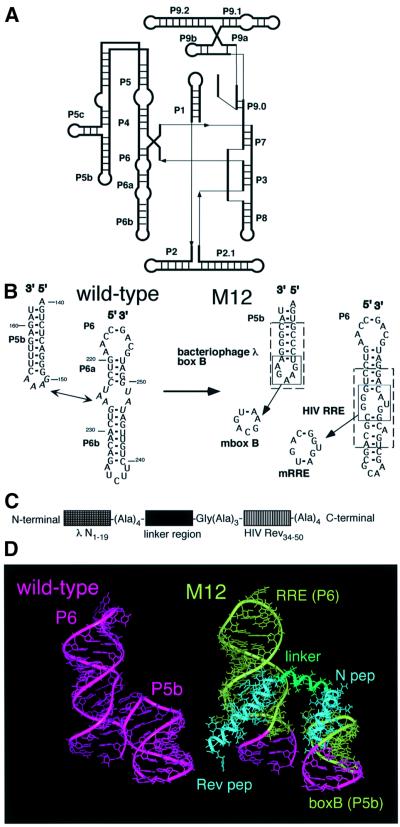
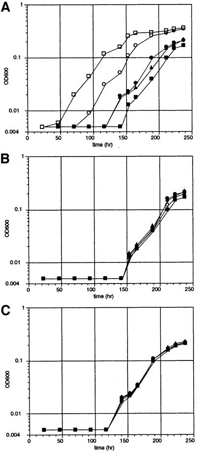
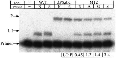
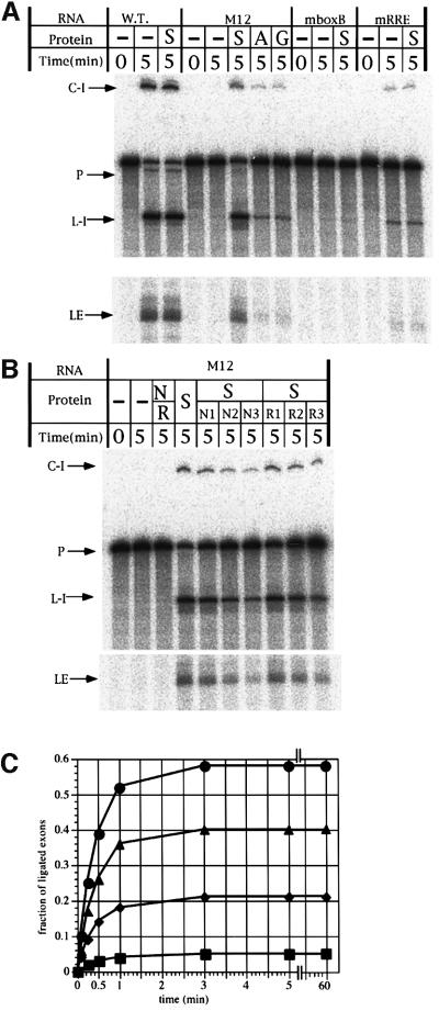
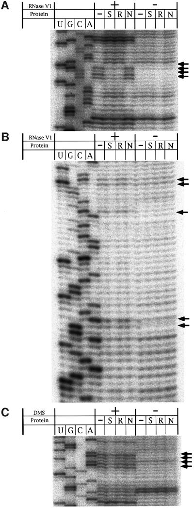
Ribonucleoproteins (RNPs) consisting of derivatives of a ribozyme and an RNA-binding protein were designed and constructed based upon high-resolution structures of the corresponding prototype molecules, the Tetrahymena group I self-splicing intron RNA and two proteins (bacteriophage lambdaN and HIV Rev proteins) containing RNA-binding motifs. The splicing reaction proceeds efficiently only when the designed RNA associates with the designed protein either in vivo or in vitro. In vivo mutagenic protein selection was effective for improving the capability of the protein. Kinetic analyses indicate that the protein promotes RNA folding to establish an active conformation. The fact that the conversion of a ribozyme to an RNP can be accomplished by simple molecular design supports the RNA world hypothesis and suggests that a natural active RNP might have evolved readily from a ribozyme.


Figures

None
Fig. 1. Design of the RNP. (A) Schematic representation of the secondary structure of the Tetrahymena ribozyme. (B) Secondary structures of P5b and P6 domains of the Tetrahymena ribozyme and its derivative (M12) used in this study. Nucleotides involved in the RNA–RNA interactions are italicized. Bacteriophage λ boxB and HIV RRE in M12 RNA are boxed by dotted lines. The mutants (mboxB and mRRE) lacking one of the binding sites are also indicated. (C) Schematic representation of the protein employed in this study. (D) Three-dimensional model of the RNP consisting of M12 RNA and the designed protein. The linker region of the protein is represented by a tentatively inserted 12mer α-helix (green).
None
Fig. 2. The in vitro splicing reaction of the designed RNP. (A) Schematic representation of the proteins employed in this study. Mutations in RNA binding sites of pep AmN and pep AmRev are also indicated using the one-letter code. (B) An autoradiogram is shown for the splicing reactions with 10 nM 32P-labeled precursor RNA in the presence or absence of 3 µM protein. G, pep G; A, pep A; N, pep AmN; R, pep AmRev; D, pep Δlinker; C-I, circular form of spliced intron; L-I, linear form of spliced intron; LE, ligated exons.
None
Fig. 3. Time-courses for the growth of E.coli cells containing the derivative of the Tetrahymena intron ribozyme and the activator protein. Cells were incubated in M9 medium containing lactose at 37°C. (A) Time-courses for cells containing M12 plasmid transformed with N pep (closed squares), pep A (closed triangles), pep G (closed circles) and pep S (open circles) plasmids and cells containing wild-type plasmid transformed with N pep plasmid (open squares). (B) Time-courses for cells containing the mboxB plasmid transformed with the respective proteins described for (A). (C) Time-courses for cells containing the mRRE plasmid transformed with the respective proteins as described for (A).
None
Fig. 4. Primer extension analysis of the precursor and intron. Ribozymes and proteins were overexpressed for 10 min prior to the isolation of the total RNA. P, cDNA from the RNA containing an intact 5′ splice site; L-I, cDNA from linear intron RNA; Primer, unreacted primer; WT, wild-type Tetrahymena ribozyme; ΔP5abc, ΔP5abc ribozyme; N, N pep; S, pep S; A, pep A; G, pep G.
None
Fig. 5. The splicing reaction with the selected protein in vitro. (A) An autoradiogram is shown for the splicing reactions with 10 nM 32P-labeled precursor RNA in the presence or absence of 100 nM protein. WT, wild-type Tetrahymena ribozyme; S, pep S; A, pep A; G, pep G; C-I, circular form of spliced intron; L-I, linear form of spliced intron; LE, ligated exons. (B) An autoradiogram is shown for the splicing reactions with 10 nM 32P-labeled precursor RNA in the presence or absence of various concentrations of proteins. Lane S, 100 nM pep S; lane N/R, 1 µM pep AmN plus 1 µM pep AmRev; lanes S/N1, S/N2, S/N3, 100 nM pep S plus 100, 200 or 400 nM pep AmN, respectively; lanes S/R1, S/R2, S/R3, 100 nM pep S plus 100, 200 or 400 nM pep AmRev, respectively. (C) Time-courses of the splicing reactions with 10 nM 32P-labeled M12 precursor RNA in the presence or absence (squares) of 3 µM pep S (circles), pep A (triangles) or pep G (diamonds).
None
Fig. 6. RNase and chemical footprints of M12 in the presence or absence of the designed proteins. Protection studies were carried out with RNase V1 or DMS. Accessibility was monitored by primer extension using RNase- or DMS-treated samples as templates. Autoradiograms are shown for RNase footprints of box B (A) and RRE (B) region and chemical footprints of the A-rich bulge (C). Arrows indicate sites that show protection from RNase or DMS.
None
Fig. 7. The splicing reaction of the designed RNP was tested in the presence of various concentrations of protein. The final extent of the splicing reaction of M12 with pep A, pep G or pep S is shown with closed triangles, closed circles and open circles, respectively, and that of the wild type or M12 is shown with dotted lines.

References

    1. Battiste J.L., Mao,H., Rao,N.S., Tan,R., Muhandiram,D.R., Kay,L.E., Frankel,A.D. and Williamson,J.R. (1996) α helix–RNA major groove recognition in an HIV-1 rev peptide–RRE RNA complex. Science, 273, 1547–1551. - PubMed
    1. Burke J.M. (1996) Structural analysis and modifications of the hairpin ribozyme. In Eckstein,F. and Lilley,D.M.J. (eds), Catalytic RNA. Springer, Berlin, Germany, pp. 129–143.
    1. Campisi D.M., Calabro,V. and Frankel,A.D. (2001) Structure-based design of a dimeric RNA–peptide complex. EMBO J., 20, 178–186. - PMC - PubMed
    1. Caprara M.G., Mohr,G. and Lambowitz,A.M. (1996) A tyrosyl-tRNA synthetase protein induces tertiary folding of the group I intron catalytic core. J. Mol. Biol., 257, 512–531. - PubMed
    1. Cate J.H., Gooding,A.R., Podell,E., Zhou,K., Golden,B.L., Kundrot,C.E., Cech,T.R. and Doudna,J.A. (1996a) Crystal structure of a group I ribozyme domain: principles of RNA packing. Science, 273, 1678–1685. - PubMed

Publication types

MeSH terms