. 2001 Oct 1;29(19):3997-4005.
doi: 10.1093/nar/29.19.3997.

Methylation of the nucleobases in RNA oligonucleotides mediates duplex-hairpin conversion

Affiliations

Methylation of the nucleobases in RNA oligonucleotides mediates duplex-hairpin conversion

R Micura et al. Nucleic Acids Res. .

Abstract

We have systematically investigated the duplex to hairpin conversion of oligoribonucleotides under the aspect of nucleobase methylation. The first part of our study refers to the self-complementary sequence rCGCGAAUUCGCGA, which forms a stable Watson-Crick base paired duplex under various buffer conditions. It is shown that this sequence is forced to adopt a hairpin conformation if one of the central 6 nt is replaced by the corresponding methylated nucleotide, such as 1-methylguanosine N(2),N(2)-dimethylguanosine, N(6),N(6)-dimethyladenosine (m(6)(2)A) or 3-methyluridine. On the other hand, the duplex structure is retained and even stabilized by replacement of a central nucleotide with N(2)-methylguanosine (m(2)G) or N(4)-methylcytidine. A borderline case is represented by N(6)-methyladenosine (m(6)A). Although generally a duplex-preserving modification, our data indicate that m(6)A in specific strand positions and at low strand concentrations is able to effectuate duplex-hairpin conversion. Our studies also include the ssu ribosomal helix 45 sequence motif, rGACCm(2)GGm(6)(2)Am(6)(2)AGGUC. In analogy, it is demonstrated that the tandem m(6)(2)A nucleobases of this oligoribonucleotide prevent duplex formation with complementary strands. Therefore, it can be concluded that nucleobase methylations at the Watson-Crick base pairing site provide the potential not only to modulate but to substantially affect RNA structure by formation of different secondary structure motifs.


Figures

None
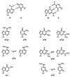
Scheme 1. Site-specific methylation of a palindromic duplex. M, N, nucleotides capable of complementary Watson–Crick base pairing; mN, mM, methylated nucleotides. Depending on the kind of methylation the duplex is either converted into a hairpin (A) or the duplex is retained (B). The bold numbers refer to the sequences investigated (compare Table 1).
Figure 1
Figure 1
Watson–Crick base pairing pattern of adenine-uracil (A-U) and guanine-cytosine (G-C) (above). Chemical constitution and conformation of methylated nucleotides encountered in cellular RNAs: 1-methylguanosine (m1G), N2,N2-dimethylguanosine (m22G), N6,N6-dimethyladenosine (m62A), 3-methyluridine (m3U), 1-methylinosine (m1I), N2-methylguanosine (m2G), N6-methyladenosine (m6A) and N4-methylcytidine (m4C). Preferred conformations of m4C, m6A and m2G are according to Engel and von Hippel (43).
Figure 2
Figure 2
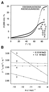
(A) Melting profiles of rCGCGAAUUCGCGA 3 and rCGCm1GAAUUCGCGA 4 (3 µM, 1.0 M NaCl, 10 mM Na2HPO4, pH 7.0). (B) Dependence of melting transitions on strand concentration c of sequences 3 and 4, (0.15 M or 1.0 M NaCl, 10 mM Na2HPO4, pH 7.0): 1/T versus lnc plots. H, hyperchromicity; T, temperature.
Figure 3
Figure 3
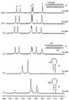
Selected 1H NMR (500 MHz) NH spectra: rCGCGAAUUCGCGA 3 (duplex, assignment according to 42), rCGCm2GAAUUCGCGA 5 (duplex), rCGCm1GAAUUCGCGA 4 (hairpin) and rCGCm22GAAUUCGCGA 6 (hairpin, asterisks indicate signals for competitive duplex formation to a minor extent). Total strand concentrations are as indicated. H2O/D2O, 9:1; sodium arsenate buffer, pH 7.4; 25 mM for 3, 5 and 6, 12.5 mM for 4)
Figure 4
Figure 4
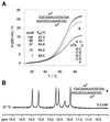
(A) Examples of UV melting curves for the sequence rCGCGm6AAUUCGCGA 9. Strand concentrations <5 µM result in low hyperchromicites and invariable melting transitions of Tm = 69.5°C, indicating hairpin formation. Higher concentrations yield increased hyperchromicities and a linear 1/T versus lnc correlation (37–313 µM, Tm = 64.2–69.2°C), indicating duplex formation. 150 mM NaCl, 10 mM Na2HPO4, pH 7.0. (B) 1H NMR (500 MHz) NH spectrum of rCGCGm6AAUUCGCGA 9 at a concentration of 0.2 mM corroborates formation of a duplex (27°C, 25 mM arsenate buffer, pH 7.4).
Figure 5
Figure 5
UV melting curves of rGACCGGAAGGUC/GACCUUCCGGUC 20 and rGACCm2GGm62Am62AGGUC/GACCUUCCGGUC 21. c = 3 µM. 150 mM NaCl, 10 mM Na2HPO4, pH 7.0.

Similar articles

Cited by

References

    1. Crain P.F. and McCloskey,J.A. (1997) The RNA modification database. Nucleic Acids Res., 25, 126–127. - PMC - PubMed
    1. Björk G.R. (1995) Biosynthesis and function of modified nucleosides. In Söll,D. and RajBhandary,U. (eds), tRNA: Structure, Biosynthesis and Function. ASM Press, Washington, DC, pp. 165–205.
    1. Yokoyama S. and Nishimura,S. (1995) Modified nucleosides and codon recognition. In Söll,D. and RajBhandary,U. (eds), tRNA: Structure, Biosynthesis and Function. ASM Press, Washington, DC, pp. 207–223.
    1. Motorin Y. and Grosjean,H. (1998) Appendix 1: Chemical structures and classification of post-transcriptionally modified nucleosides in RNA. In Grosjean,H. and Benne,R. (eds), Modification and Editing of RNA. ASM Press, Washington, DC, pp. 543–549.
    1. Grosjean H. and Benne,R. (eds) (1998) Modification and Editing of RNA. ASM Press, Washington, DC.

Publication types

MeSH terms