. 2001 Oct 1;29(19):4043-51.
doi: 10.1093/nar/29.19.4043.

Specific distribution of the Saccharomyces cerevisiae linker histone homolog HHO1p in the chromatin

Affiliations

Specific distribution of the Saccharomyces cerevisiae linker histone homolog HHO1p in the chromatin

I Freidkin et al. Nucleic Acids Res. .

Abstract

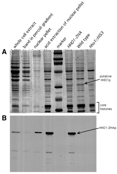
In virtually all eukaryotic organisms, linker DNA between nucleosomes is associated with a histone termed linker histone or histone H1. In Saccharomyces cerevisiae, HHO1 encodes a putative linker histone with very significant homology to histone H1. The encoded protein is expressed in the nucleus, but has not been shown to affect global chromatin structure, nor has its deletion shown any detectable phenotype. In vitro chromatin assembly experiments with recombinant HHO1p have shown that it is able to complex with dinuncleosomes in a similar manner to histone H1. Here we report that while disruption of HHO1 has little affect on RNA levels of most cellular transcripts, there are numerous exceptions. Measurement of HHO1p concentration in the wild-type cell showed a stoichiometry of about one HHO1p molecule per 37 nucleosomes. Localization of HHO1p in the chromatin, using an immunoprecipitation technique, showed preferential HHO1p binding to rDNA sequences. These results suggest that HHO1p may play a similar role to linker histones, but at restricted locations in the chromatin.


Figures

Figure 1
Figure 1
Histone preparations from yeast nuclei. (A) Coomassie staining of fractions from various stages of purification. Lane 1, whole cell extract; lane 2, band in Percoll gradient; lane 3, nuclear pellet; lane 4, acid extraction of nuclear pellet; lane 5, marker (mol. wt 220, 97.4, 66, 46, 30 21.5 and 14.3 kDa, respectively); lane 6, HHO1–2HA; lane 7, wild-type; lane 8, hho1::HIS3. The putative HHO1p band is present in the wild-type but not in hho1::HIS3 and has a higher molecular weight in the HHO1–2HA lane. (B) Western analysis of (A) with anti-HA antibody.
Figure 2
Figure 2
Northern analysis of whole cell RNA from strains that were wild-type for HHO1 (WT) or were hho1Δ (Δ). Each filter was probed with a radiolabeled PCR fragment containing sequences from the gene listed above each panel. Below the panel is an ethidium stain of the 18S and 28S rRNAs in each sample showing that equal amounts of RNA were loaded in each lane.
Figure 3
Figure 3
Quantitation of HHO1p in the cell. Western analysis was performed comparing known amounts of recombinant GST–HHO1p–2HA (58 kDa) with unknown quantities of HHO1p–2HA (30 kDa). Lanes 1–7 contain 37.5, 18.7, 9.4, 4.7, 2.3, 1.2 and 0.6 ng GST–HHO1p–2HA, respectively. Lanes 8–14 contain whole cell lysate from 13.2 × 10⁷, 6.6 × 10⁷, 3.3 × 10⁷, 1.6 × 10⁷, 0.8 × 10⁷ and 0.4 × 10⁷ cells, respectively.
Figure 4
Figure 4
Immunoprecipitation of HHO1p–2HA-associated chromatin. (A) Ethidium stained agarose gel of restriction digested whole cell DNA. M, λ Eco130I + MluI digested DNA marker (sizes in bp are to left of figure). Lane 1, EcoRI; lane 2, HindIII; lane 3, BglII; lane 4, XhoI. Repeated DNA sequences can be seen as bands on the gel. (B) Southern blot of left panel using immunoprecipitated DNA as probe. All the bands that appear were identified in Foley et al. (41) as encoding rRNA. No other repeated sequences were detected. (C) (Left) rDNA and CUP1 sequences (primers IF9–IF14) were multiplex PCR amplified from total genomic and immunoprecipitated DNA as described in Materials and Methods. The sizes of the amplicons are listed in parentheses. Lane 1, total genomic DNA; lane 2, immunoprecipated DNA. Duplicate lanes are duplicate PCR reactions. Band intensities are quantified in Table 2. (Right) LYS9, CTR1, SAG1, FET3 and FRE1 (primers IF27–IF34) were PCR amplified as in the left panel. All these genes are single copy in the genome. Neither rDNA nor CUP1 were included in the right panel as they are in multiple copies in the genome and they overwhelm the PCR reaction when amplified in the same tube. While none of the DNA fragments in the right panel is specifically immunoprecipitated one relative to the other, it is evident in the left panel that only the rDNA sequences are preferentially immunoprecipitated. About 10-fold more DNA template was used in the multiplex PCR reaction for the single copy genes (right) as for the multicopy genes (left).

References

    1. Van Holde K.E. (1989) Chromatin. Springer-Verlag, New York, NY.
    1. Kornberg R.D. (1974) Chromatin structure: a repeating unit of histones and DNA. Science, 184, 868–871. - PubMed
    1. Kornberg R.D. and Thomas,J.O. (1974) Chromatin structure; oligomers of the histones. Science, 184, 865–868. - PubMed
    1. Graziano V., Gerchman,S.E., Schneider,D.K. and Ramakrishnan,V. (1994) Histone H1 is located in the interior of the chromatin 30-nm filament. Nature, 368, 351–354. - PubMed
    1. Doenecke D., Tonjes,R. and Kress,H. (1988) The H1 and core histone subtypes: differential gene expression and varied primary structures. Adv. Enzyme Regul., 27, 107–120. - PubMed

Publication types