

Changes in gene expression in macrophages infected with Mycobacterium tuberculosis: a combined transcriptomic and proteomic approach
- PMID: 11576227
- PMCID: PMC1783284
- DOI: 10.1046/j.0019-2805.2001.01274.x
Changes in gene expression in macrophages infected with Mycobacterium tuberculosis: a combined transcriptomic and proteomic approach
Abstract
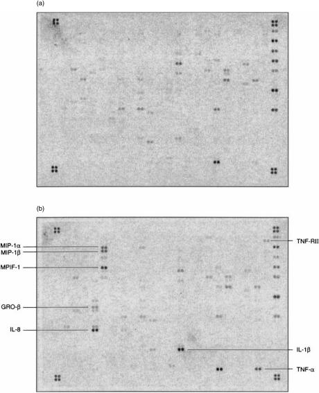
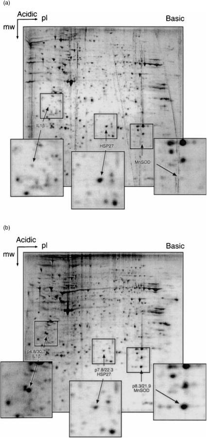
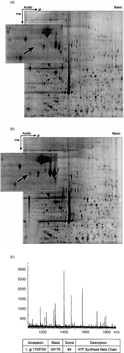
We investigated the changes which occur in gene expression in the human macrophage cell line, THP1, at 1, 6 and 12 hr following infection with Mycobacterium tuberculosis. The analysis was carried out at the transcriptome level, using microarrays consisting of 375 human genes generally thought to be involved in immunoregulation, and at the proteomic level, using two-dimensional gel electrophoresis and mass spectrometry. The analysis of the transcriptome using microarrays revealed that many genes were up-regulated at 6 and 12 hr. Most of these genes encoded proteins involved in cell migration and homing, including the chemokines interleukin (IL)-8, osteopontin, monocyte chemotactic protein-1 (MCP-1), macrophage inflammatory protein-1alpha (MIP-1alpha), regulated on activation, normal, T-cell expressed and secreted (RANTES), MIP-1beta, MIP-3alpha, myeloid progenitor inhibitory factor-1 (MPIF-1), pulmonary and activation regulated chemokine (PARC), growth regulated gene-beta (GRO-beta), GRO-gamma, MCP-2, I-309, and the T helper 2 (Th2) and eosinophil-attracting chemokine, eotaxin. Other genes involved in cell migration which were up-regulated included the matrix metalloproteinase MMP-9, vascular endothelial growth factor (VEGF) and its receptor Flk-1, the chemokine receptor CCR3, and the cell adhesion molecules vesicular cell adhesion molecule-1 (VCAM-1) and integrin a3. In addition to the chemokine response, genes encoding the proinflammatory cytokines IL-1beta (showing a 433-fold induction), IL-2 and tumour necrosis factor-alpha (TNF-alpha), were also found to be induced at 6 and/or 12 hr. It was more difficult to detect changes using the proteomic approach. Nevertheless, IL-1beta was again shown to be strongly up-regulated. The enzyme manganese superoxide dismutase was also found to be strongly up-regulated; this enzyme was found to be macrophage-, rather than M. tuberculosis, derived. The heat-shock protein hsp27 was found to be down-regulated following infection. We also identified a mycobacterial protein, the product of the atpD gene (thought to be involved in the regulation of cytoplasmic pH) in the infected macrophage extracts.
Figures
Similar articles
-
Cytokine and Chemokine Concentration in the Tear of Patients with Age-Related Cataract.Curr Eye Res. 2020 Sep;45(9):1101-1106. doi: 10.1080/02713683.2020.1715445. Epub 2020 Jan 24. Curr Eye Res. 2020. PMID: 31928443
-
Gene expression profiling of human macrophages at late time of infection with Mycobacterium tuberculosis.Immunology. 2006 Aug;118(4):449-60. doi: 10.1111/j.1365-2567.2006.02378.x. Immunology. 2006. PMID: 16895554 Free PMC article.
-
Differential chemokine response of murine macrophages stimulated with cytokines and infected with Listeria monocytogenes.Int Immunol. 1998 Jun;10(6):757-65. doi: 10.1093/intimm/10.6.757. Int Immunol. 1998. PMID: 9678756
-
Infection of human macrophages and dendritic cells with Mycobacterium tuberculosis induces a differential cytokine gene expression that modulates T cell response.J Immunol. 2001 Jun 15;166(12):7033-41. doi: 10.4049/jimmunol.166.12.7033. J Immunol. 2001. PMID: 11390447
-
Redirection of doublecortin-positive cell migration by over-expression of the chemokines MCP-1, MIP-1α and GRO-α in the adult rat brain.Neuroscience. 2014 Feb 28;260:240-8. doi: 10.1016/j.neuroscience.2013.12.022. Epub 2013 Dec 18. Neuroscience. 2014. PMID: 24361178
Cited by
-
IP-10, MCP-1, MCP-2, MCP-3, and IL-1RA hold promise as biomarkers for infection with M. tuberculosis in a whole blood based T-cell assay.BMC Res Notes. 2009 Feb 4;2:19. doi: 10.1186/1756-0500-2-19. BMC Res Notes. 2009. PMID: 19193208 Free PMC article.
-
Caseation of human tuberculosis granulomas correlates with elevated host lipid metabolism.EMBO Mol Med. 2010 Jul;2(7):258-74. doi: 10.1002/emmm.201000079. EMBO Mol Med. 2010. PMID: 20597103 Free PMC article.
-
Use of an Integrated Multi-Omics Approach To Identify Molecular Mechanisms and Critical Factors Involved in the Pathogenesis of Leptospira.Microbiol Spectr. 2023 Feb 28;11(2):e0313522. doi: 10.1128/spectrum.03135-22. Online ahead of print. Microbiol Spectr. 2023. PMID: 36853003 Free PMC article.
-
Mycobacterium tuberculosis Requires Cholesterol Oxidase to Disrupt TLR2 Signalling in Human Macrophages.Mediators Inflamm. 2019 Dec 1;2019:2373791. doi: 10.1155/2019/2373791. eCollection 2019. Mediators Inflamm. 2019. PMID: 31871425 Free PMC article.
-
System-wide coordinates of higher order functions in host-pathogen environment upon Mycobacterium tuberculosis infection.Sci Rep. 2018 Mar 22;8(1):5079. doi: 10.1038/s41598-018-22884-8. Sci Rep. 2018. PMID: 29567998 Free PMC article.
References
-
- Brightbill HD, Libraty DH, Krutzik SR, et al. Host defense mechanisms triggered by microbial lipoproteins through toll-like receptors. Science. 1999;285:732–6. 10.1126/science.285.5428.732. - DOI - PubMed
-
- Means TK, Wang S, Lien E, Yoshimura A, Golenbock DT, Fenton MJ. Human toll-like receptors mediate cellular activation by Mycobacterium tuberculosis. J Immunol. 1999;163:3920–7. - PubMed
-
- Hirsch CS, Ellner JJ, Russell DG, Rich EA. Complement receptor-mediated uptake and tumor necrosis factor-alpha-mediated growth inhibition of Mycobacterium tuberculosis by human alveolar macrophages. J Immunol. 1994;152:743–53. - PubMed
-
- Schlesinger LS. Macrophage phagocytosis of virulent but not attenuated strains of Mycobacterium tuberculosis is mediated by mannose receptors in addition to complement receptors. J Immunol. 1993;150:2920–30. - PubMed
Publication types
MeSH terms
Substances
Grants and funding
LinkOut - more resources
Full Text Sources
Other Literature Sources
Medical
Research Materials
Miscellaneous
