

Concomitant progressive multifocal leucoencephalopathy and primary central nervous system lymphoma expressing JC virus oncogenic protein, large T antigen
- PMID: 11577180
- PMCID: PMC1187095
- DOI: 10.1136/mp.54.5.354
Concomitant progressive multifocal leucoencephalopathy and primary central nervous system lymphoma expressing JC virus oncogenic protein, large T antigen
Abstract
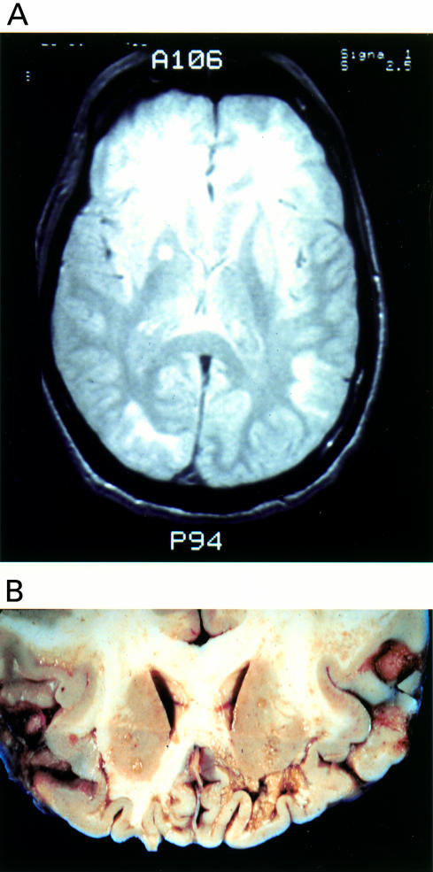
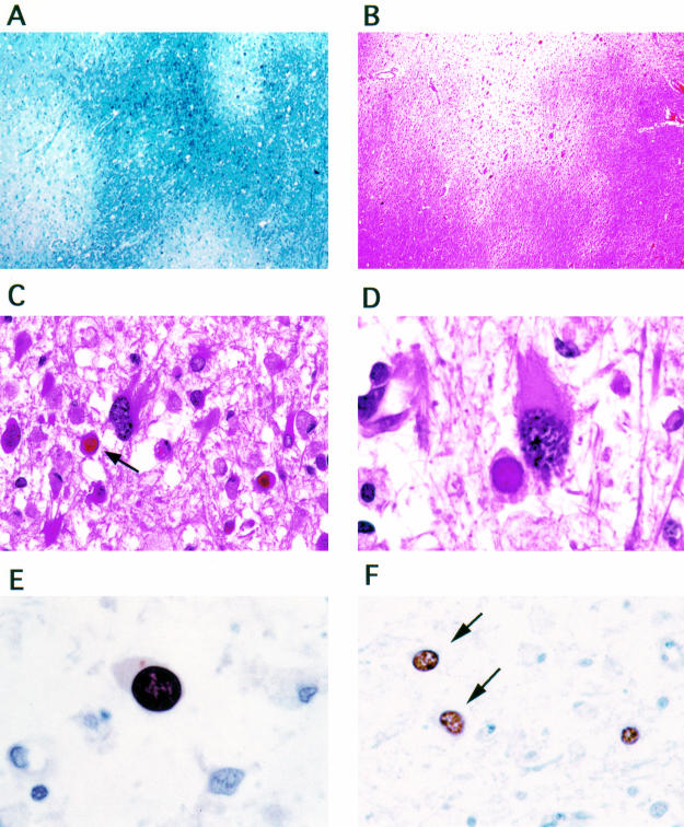
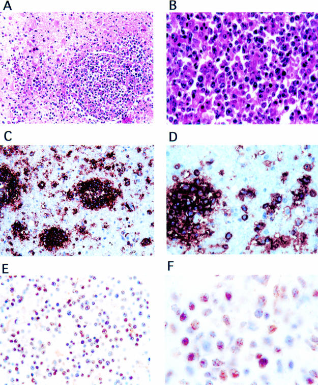
This report describes the concomitant occurrence of the JC virus (JCV) induced demyelinating disease progressive multifocal leucoencephalopathy (PML) and a primary central nervous system lymphoma (PCNS-L) in a patient with AIDS. Postmortem neuropathological examination revealed characteristic features of PML including multiple lesions of demyelination, enlarged oligodendrocytes with hyperchromatic nuclei (many containing eosinophilic intranuclear inclusions), and enlarged astrocytes with bizarre hyperchromatic nuclei. Immunohistochemical analysis demonstrated the expression of the JCV capsid protein VP-1 in the nuclei of infected oligodendrocytes and astrocytes. The PCNS-L lesion located in the basal ganglia was highly cellular, distributed perivascularly, and consisted of large atypical plasmacytoid lymphocytes. Immunohistochemical examination of this neoplasm identified it to be of B cell origin. Moreover, expression of the JCV oncogenic protein, T antigen, was detected in the nuclei of the neoplastic lymphocytes. This study provides the first evidence for a possible association between JCV and PCNS-L.
Figures
Similar articles
-
Detection of JC virus type 1 in peripheral lymphocytes, brain and cerebrospinal fluid from two Korean AIDS patients with progressive multifocal leukoencephalopathy.Intervirology. 2002;45(2):94-100. doi: 10.1159/000063229. Intervirology. 2002. PMID: 12145541
-
[Progressive multifocal leukoencephalopathy. Demyelinating viral disease--common complication of AIDS].Lakartidningen. 2001 Sep 26;98(39):4206-11, 4213. Lakartidningen. 2001. PMID: 11680156 Review. Swedish.
-
Evidence for involvement of transforming growth factor beta1 signaling pathway in activation of JC virus in human immunodeficiency virus 1-associated progressive multifocal leukoencephalopathy.Arch Pathol Lab Med. 2004 Mar;128(3):282-91. doi: 10.5858/2004-128-282-EFIOTG. Arch Pathol Lab Med. 2004. PMID: 14987161
-
JC virus-specific cytotoxic T lymphocytes in individuals with progressive multifocal leukoencephalopathy.J Virol. 2001 Apr;75(7):3483-7. doi: 10.1128/JVI.75.7.3483-3487.2001. J Virol. 2001. PMID: 11238876 Free PMC article.
-
[Progressive multifocal leukoencephalopathy: virological and neuropathological aspects].Arch Anat Cytol Pathol. 1997;45(2-3):127-34. Arch Anat Cytol Pathol. 1997. PMID: 9382604 Review. French.
Cited by
-
Primary central nervous system lymphoma expressing the human neurotropic polyomavirus, JC virus, genome.J Virol. 2004 Apr;78(7):3462-9. doi: 10.1128/jvi.78.7.3462-3469.2004. J Virol. 2004. PMID: 15016869 Free PMC article.
-
Diffuse large B cell lymphoma secondary to JC virus in progressive multifocal leukoencephalopathy.J Neurovirol. 2019 Dec;25(6):883-886. doi: 10.1007/s13365-019-00760-z. Epub 2019 May 28. J Neurovirol. 2019. PMID: 31140130
-
Co-Detection of EBV and Human Polyomavirus JCPyV in a Case of AIDS-Related Multifocal Primary Central Nervous System Diffuse Large B-Cell Lymphoma.Viruses. 2023 Mar 15;15(3):755. doi: 10.3390/v15030755. Viruses. 2023. PMID: 36992464 Free PMC article.
-
The Role of the JC Virus in Central Nervous System Tumorigenesis.Int J Mol Sci. 2020 Aug 28;21(17):6236. doi: 10.3390/ijms21176236. Int J Mol Sci. 2020. PMID: 32872288 Free PMC article. Review.
-
Case Report: Findings Suggestive of Paraclinical Progressive Multifocal Leukoencephalopathy and Lung Cancer-Derived Brain Metastases in an MS Patient Treated With Fingolimod.Front Neurol. 2021 Feb 3;12:561158. doi: 10.3389/fneur.2021.561158. eCollection 2021. Front Neurol. 2021. PMID: 33613428 Free PMC article.
References
-
- Padgett BL, Walker DL. Prevalence of antibodies in human sera against JC virus, an isolate from a case of progressive multifocal leukoencephalopathy. J Infect Dis 1973;127:467–70. - PubMed
-
- Taguchi F, Kajioka J, Miyamura T. Prevalence rate and age of acquisition of antibodies against JC virus and BK virus in human sera. Microbiol Immunol 1982;26:1057–64. - PubMed
-
- Berger JR, Concha M. Progressive multifocal leukoencephalopathy: the evolution of a disease once considered rare. J NeuroVirol 1995;5:5–18. - PubMed
-
- Berger JR, Pall L, Lanska D, et al. Progressive multifocal leukoencephalopathy in patients with HIV infection. J Neurovirol 1998;4:59–68. - PubMed
Publication types
MeSH terms
Substances
LinkOut - more resources
Full Text Sources
Medical