. 2001 Nov-Dec;39(5-6):341-8.
doi: 10.1016/s0197-0186(01)00041-9.

Interleukin-1 promotion of MAPK-p38 overexpression in experimental animals and in Alzheimer's disease: potential significance for tau protein phosphorylation

Affiliations

Interleukin-1 promotion of MAPK-p38 overexpression in experimental animals and in Alzheimer's disease: potential significance for tau protein phosphorylation

J G Sheng et al. Neurochem Int. 2001 Nov-Dec.

Abstract

Activated (phosphorylated) mitogen-activated protein kinase p38 (MAPK-p38) and interleukin-1 (IL-1) have both been implicated in the hyperphosphorylation of tau, a major component of the neurofibrillary tangles in Alzheimer's disease. This, together with findings showing that IL-1 activates MAPK-p38 in vitro and is markedly overexpressed in Alzheimer brain, suggest a role for IL-1-induced MAPK-p38 activation in the genesis of neurofibrillary pathology in Alzheimer's disease. We found frequent colocalization of hyperphosphorylated tau protein (AT8 antibody) and activated MAPK-p38 in neurons and in dystrophic neurites in Alzheimer brain, and frequent association of these structures with activated microglia overexpressing IL-1. Tissue levels of IL-1 mRNA as well as of both phosphorylated and non-phosphorylated isoforms of tau were elevated in these brains. Significant correlations were found between the numbers of AT8- and MAPK-p38-immunoreactive neurons, and between the numbers of activated microglia overexpressing IL-1 and the numbers of both AT8- and MAPK-p38-immunoreactive neurons. Furthermore, rats bearing IL-1-impregnated pellets showed a six- to seven-fold increase in the levels of MAPK-p38 mRNA, compared with rats with vehicle-only pellets (P<0.0001). These results suggest that microglial activation and IL-1 overexpression are part of a feedback cascade in which MAPK-p38 overexpression and activation leads to tau hyperphosphorylation and neurofibrillary pathology in Alzheimer's disease.


Figures

Fig. 1
Fig. 1
A. Numbers of IL-1-immunoreactive microglia, of activated-MAPK-p38-immunoreactive neurons, and of hyperphosphorylated tau (AT8)-immunoreactive neurons in Alzheimer (AD) and control patients. B. Percent of neurons immunoreactive only for activated-MAPK-p38 (P38+), for hyperphosphorylated tau (AT8), or for both markers. Values are expressed as mean ± S.E.M. ** Value is significantly different from corresponding control value (P < 0.001).
Fig. 2
Fig. 2
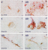
Dual-label immunohistochemistry showing morphological association of IL-1-immunoreactive microglia with neurons and neurites immunoreactive for activated MAPK-p38, and showing colocalization of activated MAPK-p38 immunoreactivity with hyperphosphorylated tau immunoreactivity within neurons in Alzheimer’s disease, (a) Control tissue showing only sparse, small microglia immunoreactive for IL-1 (brown), and no discernible immunoreactivity for activated MAPK-p38 (red), (b) Alzheimer tissue showing close association of IL-1-immunoreactive microglia (brown), with activated-MAPK-p38-immunoreactive neurites (red) in a neuritic β-amyloid plaque. Adjacent to the plaque, there are pyramidal neurons that are also immunoreactive for activated MAPK-p38 (arrows), (c) Control tissue showing weak immunoreactivity for activated MAPK-p38 (brown) and no discernible immunoreactivity for hyperphosphorylated tau (red), (d) Alzheimer tissue showing colocalization within neurons of immunoreactivity for activated MAPK-p38 (brown) and for hyperphosphorylated tau (red). Activated MAPK-p38 immunoreactivity is evident in both the cytoplasm and the nucleus, (e and f) Alzheimer tissue showing colocalization within neurites of immunoreactivity for activated MAPK-p38 (brown) and for hyperphosphorylated tau (AT8; red). Bars = 15 μm.
Fig. 3
Fig. 3
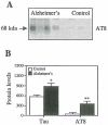
Western immunoblot analysis of total hyperphosphorylated tau (AT8) in Alzheimer and control tissue. (A) An example of Western immunoblot results from four Alzheimer and three control patients. (B) Quantification of total hyperphosphorylated tau (ATS) for 12 Alzheimer and nine control patients. Values are expressed as mean ± S.E.M. ** Value is significantly different from control at P < 0.001.
Fig. 4
Fig. 4
Reverse transcriptase-polymerase chain reaction (RT-PCR) analysis of IL-1 mRNA in Alzheimer’s disease and controls. (A) An example of RT-PCR results showing IL-1α and GAPDH mRNA levels in brain tissue from three control and four Alzheimer patients. (B) Quantification of IL-1α mRNA levels relative to GAPDH mRNA levels. Values are expressed as mean ± S.E.M. ** Value is significantly different from control (P < 0.001).
Fig. 5
Fig. 5
Linear regression analyses of correlations between (A) tissue IL-1 mRNA levels and numbers of IL-l-immunoreactive (IL-lα+) microglia; (B) numbers of IL-1+ microglia and numbers of neurons immunoreactive for activated MAPK-p38 (MAPK-p38+); (C) numbers of IL-1+ microglia and numbers of neurons immunoreactive for hyperphosphorylated tau (AT8 + ); and (D) numbers of MAPK-p38+ neurons and numbers of AT8+ neurons.
Fig. 6
Fig. 6
Reverse transcriptase-polymerase chain reaction (RT-PCR) analysis of the relative levels of MAPK-p38 mRNA in rat brain. (A) An example of RT-PCR results showing MAPK-p38 and GAPDH levels in brain from six IL-1 pellet, five sham pellets, and six unoperated-control rats. (B) Quantification of MAPK-p38 mRNA levels relative to GAPDH mRNA levels. Values are expressed as mean ± S.E.M. ** Value is significantly different from control (P < 0.0001).
Fig. 7
Fig. 7
A proposed feedback loop depicting interactions among IL-1, MAPK-p38, and tau.

References

    1. Braak H, Braak E, Strothjohann M. Abnormally phosphorylated tau protein related to the formation of neurofibrillary tangles and neuropil threads in the cerebral cortex of sheep and goat. Neurosci. Lett. 1994;171:1–4. - PubMed
    1. Drewes G, Lichtenberg-Kraag B, Döring F, et al. Mitogen activated protein (MAP) kinase transforms tau protein into an Alzheimer-like state. Eur. Mol. Biol. Org. J. 1992;11:2131–2138. - PMC - PubMed
    1. Du Y, Dodel RC, Eastwood BJ, Bales KR, Gao F, Lohmuller F, Muller U, Kurz A, Zimmer R, Evans RM, Hake A, Gasser T, Oertel WH, Griffin WST, Paul SM, Farlow MR. Association of an interleukin la polymorphism with Alzheimer’s disease. Neurology. 2000;55:480–483. - PubMed
    1. Goedert M, Jakes R, Vanmechelen E. Monoclonal antibody AT8 recognizes tau protein phosphorylated at both serine 202 and threonine 205. Neurosci. Lett. 1995;189:167–170. - PubMed
    1. Griffin WST, Stanley LC, Ling C, White L, MacLeod V, Perrot LJ, White CL, Jr, Araoz C. Brain interleukin-1 and SI00 immunoreactivity is elevated in Down syndrome and Alzheimer’s disease. Proc. Natl. Acad. Sci. USA. 1989;86:7611–7615. - PMC - PubMed

Publication types

MeSH terms