. 2001 Oct 1;536(Pt 1):35-47.
doi: 10.1111/j.1469-7793.2001.t01-1-00035.x.

Nociceptin inhibits calcium channel currents in a subpopulation of small nociceptive trigeminal ganglion neurons in mouse

Affiliations

Nociceptin inhibits calcium channel currents in a subpopulation of small nociceptive trigeminal ganglion neurons in mouse

S L Borgland et al. J Physiol. .

Abstract

1. The effects of nociceptin/orphanin FQ (N/OFQ) and opioid receptor agonists on voltage-activated calcium channel currents (I(Ca)) were examined in acutely isolated mouse trigeminal ganglion neurons using whole-cell patch-clamp recordings. These effects were correlated with responses of the neurons to capsaicin and binding of Bandeiraea simplicifolia isolectin B4 (IB4). 2. Trigeminal neurons were divided into two populations based on the presence (type 2) or absence (type 1) of a prominent T-type I(Ca). N/OFQ potently (EC(50) of 19 nM) inhibited high-voltage-activated (HVA) I(Ca) in most (82 %) small (capacitance < 12 pF) type 1 neurons, but few (9 %) larger (> 12 pF) type 1 neurons. N/OFQ inhibited I(Ca) in few (23 %) type 2 cells, and did not affect the T-type I(Ca) in any cell. 3. The mu-opioid agonists DAMGO and morphine inhibited I(Ca) in most type 1 neurons, more often (95 % versus 77 %) in the small cells. The inhibition of I(Ca) by DAMGO and morphine was more efficacious in small versus large type 1 neurons. mu-Opioids did not inhibit I(Ca) in type 2 neurons. 4. Most small type 1 neurons were sensitive to capsaicin (93 %) and bound IB4 (86 %). Fewer larger type 1 neurons responded to capsaicin (30 %) or bound IB4 (58 %). Type 2 neurons did not respond to capsaicin, although some bound IB4 (35 %). 5. Thus, N/OFQ preferentially inhibits HVA I(Ca) in a subpopulation of small nociceptive trigeminal ganglion neurons that is also highly sensitive to mu-opioid agonists.


Figures
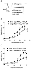
Figure 6
Figure 6. Small type 1 neurons are more sensitive to μ-opioids than large type 1 neurons
A, an example trace of HVA ICa inhibited by morphine in a type 1 neuron. Inhibition was blocked in the presence of the μ-opioid antagonist CTAP. B, DAMGO inhibited HVA ICa more potently and with greater maximal effect in small versus large type 1 neurons (at least 6 cells per point; ANOVA, P < 0.05). C, morphine inhibited HVA ICa with greater maximal effect in small versus large type 1 neurons (n = 5–8 cells per point, ANOVA, P < 0.02).
Figure 5
Figure 5
A, a representative current trace from a type 1 neuron in the presence or absence of 1 μm capsaicin. Currents were elicited by the ramp protocol shown below the example traces. B, examples of ICa currents recorded before, during and after application of DAMGO in a type 1 neuron. C, N/OFQ inhibits HVA ICa (at −7 mV) preferentially in small (< 12 pF, open symbols) versus larger (> 12 pF, filled symbols) type 1 neurons. Each data point represents the inhibition of HVA ICa by N/OFQ (300 nm to 1 μm) in a type 1 neuron. Circles represent neurons that responded and triangles represent those that did not respond to N/OFQ (see text for definition of a positive response). D, small type 1 neurons could be distinguished from large ones on the basis of responses to N/OFQ, DAMGO, capsaicin and binding of IB4. Inhibition of HVA ICa (at −7 mV) was determined in the presence or absence of 1–3 μm DAMGO or 300 nm to 1 μm N/OFQ. IB4 sensitivity was determined using fluorescence microscopy after incubation with FITC-conjugated IB4. Bars represent percentage of cells that were sensitive to the treatment. Large type 1 neurons had significantly lower frequencies of response to N/OFQ, DAMGO, capsaicin and IB4 than small type 1 neurons. *P < 0.05, **P < 0.001 (χ2-test, small versus large type 1 neurons).
Figure 4
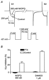
Figure 4. Inhibition of HVA ICa by N/OFQ is mediated by βγ subunits released from Gi/o proteins
A, pre-pulse relief of inhibition indicates βγ-subunit-mediated inhibition. Neurons were initially stepped from −87 to −7 mV (S1). Neurons recovered for 100 ms (approximately 80 ms has been removed for clarity, as marked by bars on the trace and voltage protocol diagram) before a conditioning pre-pulse to +123 mV for 70 ms was applied. A second test pulse (S2) was elicited by stepping the membrane potential from −87 to −7 mV. Inhibition of HVA ICa by 300 nm N/OFQ was greatly reduced in S2 as compared to S1. Leak subtraction was not used for this experiment. B, trigeminal neurons were incubated overnight at 25 °C with or without 100 ng ml−1 PTX. The PTX treatment abolished the inhibition of HVA ICa by 300 nm N/OFQ or 1 μm DAMGO. Bars represent the mean ±s.e.m. HVA ICa inhibition occurred in at least six cells per condition.
Figure 1
Figure 1. Calcium current characteristics of two populations of mouse trigeminal neurons
A and B, illustration of ICa elicited by steps from −87 to −47 and −7 mV for each cell type: type 1, neuron without T-type ICa (A); and type 2, neuron with T-type ICa (B). C, a current-voltage relationship of a neuron without LVA ICa (type 1 cell). D, a current-voltage relationship of a neuron with LVA ICa (type 2 cell). E, a frequency histogram of cell size in the two populations of neurons. Cells with LVA ICa (filled bars) are small, whereas cells without LVA ICa (open bars) have a greater range of cell sizes. F, Ni2+ inhibits LVA ICa more potently than Cd2+.
Figure 2
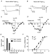
Figure 2. N/OFQ reversibly inhibits HVA ICa in sensory neurons
A, 300 nm N/OFQ reversibly inhibits HVA ICa in sensory neurons without LVA ICa (type 1). HVA ICa was elicited by stepping the membrane potential from −87 to −7 mV. B, a representative example of a current-voltage relationship of a type 1 neuron in the presence (triangles) and absence (squares) of N/OFQ (300 nm). C, N/OFQ produced a small inhibition of HVA ICa, but not LVA ICa in some neurons with ‘T-type’ICa (type 2). LVA ICa was activated by stepping membrane potential from −87 to −47 mV, HVA ICa was activated by a step to −7 mV. Approximately 60 ms of the trace, as marked by bars, has been removed for clarity. D, a current-voltage relationship of a representative type 2 neuron in the presence (triangles) or absence (squares) of 300 nm N/OFQ.
Figure 3
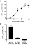
Figure 3. N/OFQ inhibits HVA ICa via activation of ORL1 receptors
A, inhibition of HVA ICa by N/OFQ is concentration dependent, with an EC50 of 19 nm. Each data point represents mean HVA ICa inhibition of at least six type 1 neurons. Error bars represent the s.e.m.B, inhibition of HVA ICa by 100 nm N/OFQ was abolished in the presence of 300 nm J113397. J113397 (300 nm) was applied to cells for 1 min before co-application of J113397 (300 nm) and N/OFQ (100 nm).
Figure 7
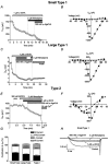
Figure 7. HVA ICa are composed of different proportions of N-type, L-type, P/Q-type and R-type in the three subpopulations of mouse sensory neurons
A, C and E, time plots of the inhibition of HVA ICa by ω-GVIA, nimodipine and ω-Aga IVA in a small type 1 neuron, and large type 1 and type 2 neurons, respectively. B, D and F, example current-voltage relationships of a small type 1, and large type 1 and type 2 neurons, respectively. Triangles represent current resistant to ω-GVIA, nimodipine and ω-Aga IVA. Squares represent current in the absence of inhibitors. G, bars represent the percentage contribution of N-type (filled bars), P/Q-type (open bars), L-type (shaded bars) and R-type ICa (hatched bars) to the peak ICa for small type 1, and large type 1 and type 2 neurons. Each proportion represents data from at least 11 cells. H, example traces from a small type 1, N/OFQ-sensitive sensory neuron showing HVA ICa following application of ω-GVIA, nimodipine and ω-Aga IVA.
Figure 8
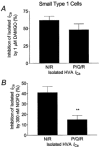
Figure 8. μ-Opioid- and ORL1-receptor agonists produce different maximal inhibitions of N/R and P/Q/R components of ICa
A, DAMGO (1 μm) produced a similar maximal inhibition of both N/R and P/Q/R ICa components. B, N/OFQ (300 nm) produced greater inhibition of N/R than P/Q/R ICa components (P < 0.01, unpaired Student's t test).

References

    1. Abdulla FA, Smith PA. Nociceptin inhibits T-type Ca2+ channel current in rat sensory neurons by a G-protein-independent mechanism. Journal of Neuroscience. 1997;17:8721–8728. - PMC - PubMed
    1. Abdulla FA, Smith PA. Axotomy reduces the effect of analgesic opioids yet increases the effect of nociceptin on dorsal root ganglion neurons. Journal of Neuroscience. 1998;18:9685–9694. - PMC - PubMed
    1. Acosta CG, Lopez HS. δ opioid receptor modulation of several voltage-dependent Ca2+ currents in rat sensory neurons. Journal of Neuroscience. 1999;19:8337–8348. - PMC - PubMed
    1. Arnot MI, Stotz SC, Jarvis SE, Zamponi GW. Differential modulation of N-type α1B and P/Q-type α1A calcium channels by different G protein α subunit isoforms. Journal of Physiology. 2000;527:203–212. - PMC - PubMed
    1. Bourinet E, Soong TW, Sutton K, Slaymaker S, Matthews E, Monteil A, Zamponi GW, Nargeot J, Snutch TP. Splicing of α1A subunit gene generates phenotypic variants of P- and Q-type calcium channels. Nature Neuroscience. 1999;2:407–415. - PubMed

Publication types

MeSH terms

LinkOut - more resources