. 2001 Oct 1;15(19):2546-61.
doi: 10.1101/gad.903101.

The neuron-specific RNA-binding protein ELAV regulates neuroglian alternative splicing in neurons and binds directly to its pre-mRNA

Affiliations

The neuron-specific RNA-binding protein ELAV regulates neuroglian alternative splicing in neurons and binds directly to its pre-mRNA

M J Lisbin et al. Genes Dev. .

Abstract

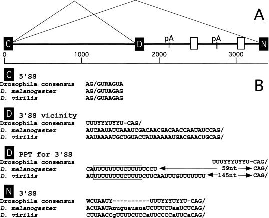
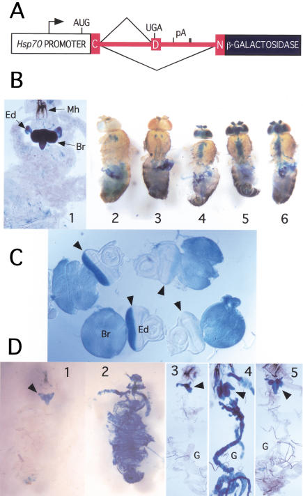
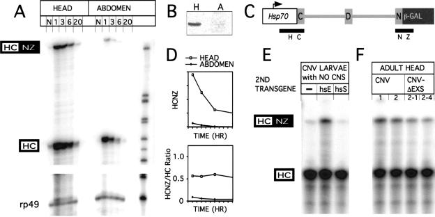
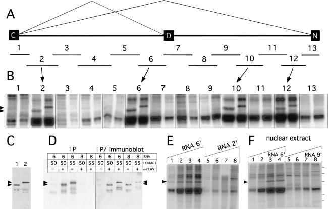
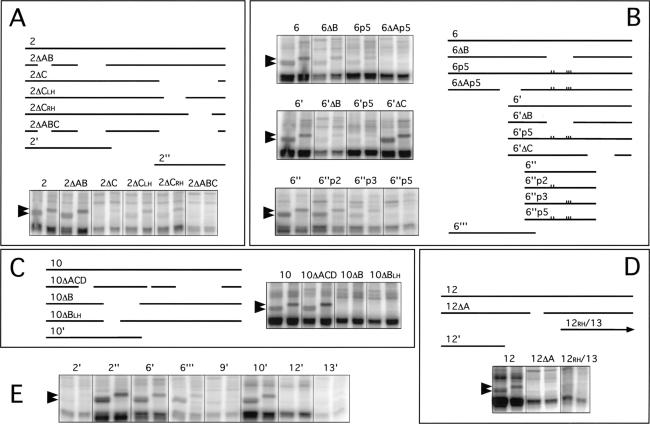
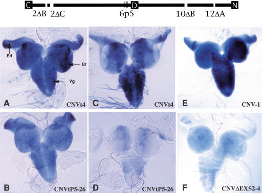
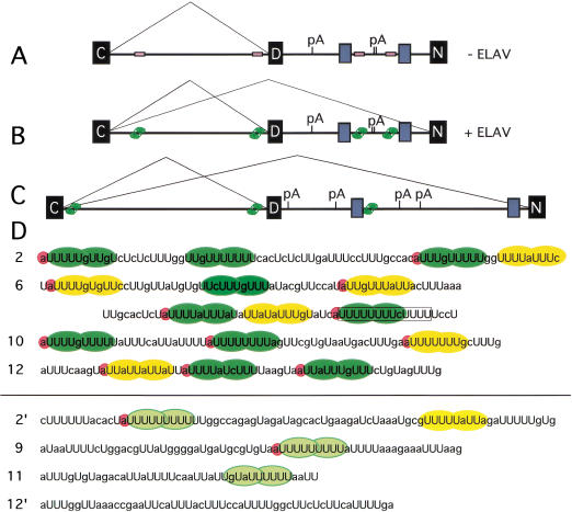
Drosophila melanogaster neural-specific protein, ELAV, has been shown to regulate the neural-specific splicing of three genes: neuroglian (nrg), erect wing, and armadillo. Alternative splicing of the nrg transcript involves alternative inclusion of a 3'-terminal exon. Here, using a minigene reporter, we show that the nrg alternatively spliced intron (nASI) has all the determinants required to recreate proper neural-specific RNA processing seen with the endogenous nrg transcript, including regulation by ELAV. An in vitro UV cross-linking assay revealed that ELAV from nuclear extracts cross-links to four distinct sites along the 3200 nucleotide long nASI; one EXS is positioned at the polypyrimidine tract of the default 3' splice site. ELAV cross-linking sites (EXSs) have in common long tracts of (U)-rich sequence rather than a precise consensus; moreover, each tract has at least two 8/10U elements; their importance is validated by mutant transgene reporter analysis. Further, we propose criteria for ELAV target sequence recognition based on the four EXSs, sites within the nASI that are (U) rich but do not cross-link with ELAV, and predicted EXSs from a phylogenetic comparison with Drosophila virilis nASI. These results suggest that ELAV regulates nrg alternative splicing by direct interaction with the nASI.


Figures

Figure 1
Figure 1
(A) Schematic of the alternatively spliced intron (nASI) of the nrg gene of D. melanogaster. C, D, and N denote the common, the default, and the neural-specific exons, respectively. The exons C, D, and N correspond to exons VI, VIIa, and VIIb of the published nrg gene (Zhao and Hortsch 1998). The nASI is flanked by the exons C and N. The default 3′SS is at position 1636 and the neural-specific 3′SS is at position 3299 of the sequence. pA indicates the positions of the three consensus polyadenylation signal sequences. The white boxes indicate blocks of homologous sequences between the two species. (B) Comparison of splice site sequences from the nASI of D. melanogaster and D. virilis. Note the conservation of the unconventional distance between the PPT, the 3′ default SS, and the 13 nt poly(U)-rich element between the two species (boxed). The default 3′SS differs considerably more from the Drosophila consensus (Mount et al. 1992) than the neural-specific 3′SS.
Figure 2
Figure 2
β-galactosidase expression from CNV minigene. (A) CNV minigene construct. The Hsp70 promoter drives the transcription of the nASI (shown in red) in frame with the downstream LacZ reporter gene. Splicing to the proximal 3′SS leads to the translation of a short peptide, whereas splicing to the distal 3′SS leads to the generation of β-galactosidase. (B) CNV expression in adults and larvae is restricted to the nervous system. CNV transgene-carrying flies or larvae were subjected to heat-shock treatment, dissected, and stained at 2 h. Variations in the treatment as noted. (1) The larval cuticle was teased apart and the entire larva was bathed in the staining solution. Note the intense staining of the brain (Br) and in the developing photoreceptors in the eye discs (Ed). Mh, mouth hooks. (2) No CNV. (3) CNV without heat shock. (4) CNV, 35°C pre heat shock only. (5) CNV, heat shock, dissected after 1 h. (6) CNV, heat shock, except 2 h at 38°C. Note that controls 2 and 3 show only the endogenous staining, but in the CNV flies after heat shock, brain, antennae, retina, and the thoracic ganglion also stain. (C) β-Galactosidase expression from the CNV is reduced in photoreceptors with reduced ELAV. Third instar CNV larval brain and eye disc stained for β-galactosidase in two genetic backgrounds for elav: elav+; CNV (wild-type ELAV level) and elave5; elavedr; CNV (reduced ELAV) expression in the photoreceptors. Each eye disc was photographed with attached brain lobe. The two left eye discs with normal ELAV levels show strong staining in the photoreceptor field of the eye disc behind the morphogenetic furrow (arrowhead) as well as staining of the attached brain lobes. Note that the right eye discs with reduced ELAV expression show a significant reduction in the staining compared to the left eye discs with wild-type ELAV expression. (D) Global expression of ELAV leads to global β-galactosidase expression from the CNV transgene. Third instar larvae were heat shocked, dissected, fixed, and stained. In panels 1 and 2, larval cuticle is cut open in the anterior and the whole animal is stained, whereas in panels 3, 4, and 5, larva is dissected and displayed with the brain attached to the mouth hooks and the digestive track (G). CNV larvae (1,3); CNV; Hsp–ELAV larvae (2,4); CNV; Hsp–Sxl (5) larvae. Note that only larvae carrying both CNV and Hsp–ELAV depict global staining (2,4).
Figure 3
Figure 3
RNA processing of the CNV transgene. (A) RNAse protection analysis of CNV transcripts. Heads and abdomens from 2- to 5-day-old CNV flies collected with no heat shock (N) or at 1-, 3-, 6-, or 20-h post 38°C heat shock. RNAse protection assay was performed as described in Materials and Methods with 10 μg of total head or abdomen RNA per lane. The antisense probe used for the protection analysis is termed HCNZ, for Hsp70 C exon/N exon lacZ (C). 321 nt are protected for a neural-specific splice, HCNZ, and 192 nt are protected for the default splice, HC (or unspliced). The rp49 probe controls for total RNA loaded. Size markers are pBR322/MspI ladder. (B) Immunoblot with α-ELAV antibodies to show ELAV level in 10 μg of total protein from adult heads, H, and abdomens, A. (D) The data from A was analyzed and quantitated on a PhosphorImager to show relative amounts of neural-processed CNV RNA. (Top graph) HCNZ-protected RNA amounts for both head and abdomen in arbitrary units. The HCNZ RNA levels decrease in the hour after heat shock. (Bottom graph) The ratio of HCNZ to HC stays relatively constant for several hours as both neural and default RNAs appear to decay with similar rates. Note: In independent experiments using an HCI probe (I, intron), the unspliced CNV, which would be seen as the HC-protected species in the figure above, is present, but in very low amounts (data not shown). (E) Ectopic expression of ELAV induces neural-specific processing of CNV transcripts in nonneuronal tissue. Third instar larvae were heat shocked, and 1 h later, the CNS was dissected out and the remaining tissue subjected to RNA extraction. RNA (5μg) was subjected to RNAse protection analysis with the HCNZ antisense probe (C). Fly heads with CNV transgene alone (−) or with Hsp–Sxl transgene (hsS) have only reduced levels of the neural-specific transcript. However, the ectopic expression of ELAV when CNV is expressed with Hsp–ELAV (hsE) leads to increased neural processing of the CNV transcripts. (F) Deletion/mutation of EXS elements within the nASI leads to a reduction in neural-specific splicing. RNA from heat-shocked CNV and CNV–ΔEXS fly heads was extracted 2 h after heat shock and used in RNAse protection analysis. The ratios of HCNZ/HC from two insertion lines of CNV (1,2) are reduced threefold in two insertion lines of the CNVΔEXS (2-1, 2-4).
Figure 4
Figure 4
UV cross-linking of ELAV from Drosophila nuclear extracts to the nASI. (A) Schematic diagram of the nASI with the 13 overlapping RNAs (1–13) used in the UV cross-linking experiment. (B) Four of the 13 RNAs UV cross-link to ELAV. [α-32P]UTP RNAs (1.2 nM) were incubated in 2 mg/mL nuclear extract with 50-kD ELAV (left) or 55-kD ELAV (right). Arrowheads indicate the two sized ELAV bands. RNAs 2, 6, 10, and 12 show cross-linking to the ELAV protein. (C) ELAV immunoblot of the two Drosophila embryonic nuclear extracts with ELAV-positive bands at 50 and 55 kD. (D) UV cross-linked ELAV is revealed by immunoprecipitation with α-ELAV in UV cross-linked extracts with RNA 6 but not with RNA 8. The two RNAs were UV cross-linked with the 50- and 55-kD nuclear extracts. Immunoprecipitates were subjected to SDS-PAGE, transferred to nitrocellulose, and exposed to PhosphorImaging plates (left panel). The filter was subsequently subjected to immunoblot analysis with α-ELAV antibodies (right panel). Note that the cross-linking bands observed with RNA 6 (left panel) correspond to the 50- and 55-kD ELAV bands (right panel). (E) UV cross-linking with increasing amounts of positive and negative ELAV cross-linking RNAs 6‘ and 2‘ (see Fig. 5E). 55-kD ELAV nuclear extract was kept constant at 1.5 mg/mL. Arrow indicates the ELAV cross-linking signal. The RNA concentrations are: (lane 1) 0.1 nM; (lane 2) 0.5 nM; (lane 3) 1.0 nM; (lane 4) 1.5 nM; (lane 5) 0.11 nM; (lane 6) 0.57 nM; (lane 7) 1.1 nM; (lane 8) 1.7 nM. (F) Titration of 55-kD ELAV nuclear extract with constant RNA concentrations. RNA 9‘ (see Fig. 5E) does not UV cross-link ELAV in embryonic nuclear extracts. RNA 6‘ (lanes 1–4) is 0.5 nM and RNA 9‘ (lanes 5–8) at 0.87 nM. Nuclear extract concentrations in mg/mL: (lanes 1 and 5) 0.5; (lanes 2 and 6) 1.0; (lanes 3 and 7) 2.0; (lanes 4 and 8) 4.0. Protein markers on the right are 90, 70, 55, 38, and 33 kD.
Figure 5
Figure 5
Deletion analysis to determine sequences responsible for ELAV UV cross-linking. Drosophila embryonic extracts were cross-linked with [α-32P]UTP nASI RNAs and processed as explained in Materials and Methods. For each RNA, deletion map schematic and dual-lane cross-linking data as in Figure 4 are shown. Deletion and point mutant sequences are given in Table 1. Arrowheads indicate the 50-KD ELAV (left lane) and 55-kD ELAV (right lane). (A) The 358 nt RNA 2 has three internal deletions, A, B, and C. RNAs deleted for C, 2ΔC, and 2ΔABC lack the ELAV cross-linking signal. Deletions of the left half 2ΔCLH or right half 2ΔCRH of C reduce but do not eliminate the ELAV signal. (B) The 319-nt RNA 6 has three internal deletions, A, B, and C, and truncated RNA 6‘, 6“‘, and 6“ have large 5′, 3′, and both 5′ and 3′ deletions, respectively. The default PPT region is deleted by ΔB and is disrupted by triple point mutant p3, whereas the nearby AU4AU3AU element is disrupted by double point mutant p2. Point mutant locations are indicated by marks on the line drawings. Data indicate the primary ELAV cross-linking site is the PPT and adjacent 5′ sequence and a suboptimal site 5′ in the ΔA region of 6“‘. (C) Four deletions were made in the 322-nt RNA 10. Deletions A, C, and D, 10ΔACD, have no effect on ELAV cross-linking; however, deletion of B, 10ΔB, and just the left half of B, 10ΔBLH, is sufficient to abolish ELAV cross-linking. (D) Deletion of part of the only significant poly(U) sequence, 12ΔA, eliminated ELAV cross-linking in the 311 nt RNA 12. (E) UV cross-linking with RNAs from A, B, C, and D with large 5′ or 3′ deletions and RNAs 9 and 13. RNA sizes in nt: (2‘) 146; (2“)112; (6‘) 198; (6“‘) 147; (9‘) 144; (10‘) 200; (12‘) 129; (13′) 106.
Figure 6
Figure 6
Representative β-galactosidase expression from mutated reporter minigenes. nASI schematic at the top shows the mutated sites in transgene CNVtp5 (6p5) and transgene CNVΔEXS (2ΔB, 2ΔC, 6p5, 10ΔB, and 12ΔA). Refer to Table 1 for sequences. C, common exon; D, default exon; N, neural-specific exon. Third instar larvae-carrying reporter minigenes were heat shocked and central nervous systems with attached eye discs were processed for X-gal staining at 4 h later (A,B,E,F) or 20 h later (C,D). The expression of CNVtp5-26 (B,D) is reduced compared to CNVt-4 (A,C). The expression of CNVΔEXS2-4 (F) is reduced compared to CNV-1 (E). Br, brain lobe; Vg, ventral ganglion; Ed, eye disc. Expression from at least two inserts was tested for each transgene.
Figure 7
Figure 7
Model for ELAV regulation of the nASI. D. melanogaster nASI (A,B) and D. virilis (C) nASI in the absence (A) and presence (B,C) of ELAV. Black boxes represent exons and gray boxes represent regions of homology between the two species. EXS sites are shown as unoccupied empty beige boxes (A) or ELAV-occupied green boxes (B,C). EXS sites in C and default splicing in neurons are assumed. pA, polyadenylation sites, are deduced from the sequence. (D) The four D. melanogaster nASI EXSs, above the line, show extended poly(U) tracts that contain multiple stretches of 8/10U (green oval shapes) that potentially accommodate ELAV RRMs 1 and 2. 5′A nucleotide associated with 8/10U stretches are highlighted in pink. The yellow ovals show blocks with only 7/10U poly(U)-rich sequence elements. The sequences below the line show nASI poly(U) elements (light green and yellow) that do not cross-link to ELAV. These contain shorter stretches that lack multiple 8/10U stretches.

References

    1. Abe R, Sakashita E, Yamamoto K, Sakamoto H. Two different RNA-binding activities for the AU-rich element and the poly(A) sequence of the mouse neuronal protein mHuC. Nucleic Acids Res. 1996;24:4895–4901. - PMC - PubMed
    1. Amara SG, Jonas V, Rosenfeld MG, Ong ES, Evans RM. Alternative RNA processing in calcitonin gene expression generates mRNAs encoding different polypeptide products. Nature. 1982;298:240–244. - PubMed
    1. Antic D, Keene JD. Embryonic lethal abnormal visual RNA-binding proteins involved in growth, differentiation, and posttranscriptional gene expression. Am J Hum Genet. 1997;61:273–278. - PMC - PubMed
    1. Antic D, Lu N, Keene JD. ELAV tumor antigen, Hel-N1, increases translation of neurofilament M mRNA and induces formation of neurites in human teratocarcinoma cells. Genes & Dev. 1999;13:449–461. - PMC - PubMed
    1. Bashaw GJ, Baker BS. The regulation of the Drosophila msl-2 gene reveals a function for Sex-lethal in translational control. Cell. 1997;89:789–798. - PubMed

Publication types

LinkOut - more resources