. 2001 Oct 1;15(19):2613-25.
doi: 10.1101/gad.915001.

LAF1, a MYB transcription activator for phytochrome A signaling

Affiliations

LAF1, a MYB transcription activator for phytochrome A signaling

M L Ballesteros et al. Genes Dev. .

Abstract

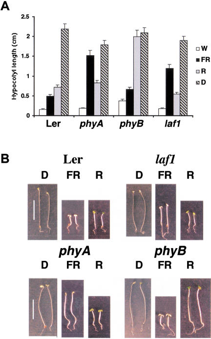
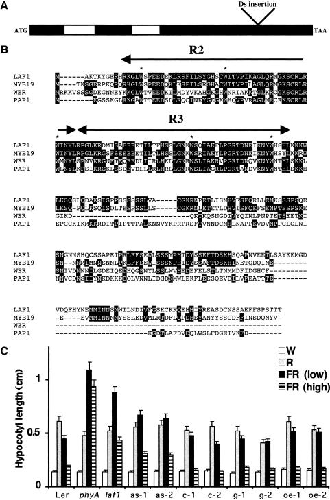
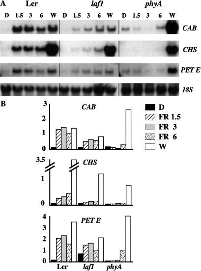
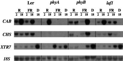
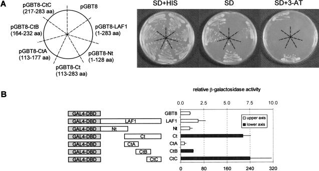
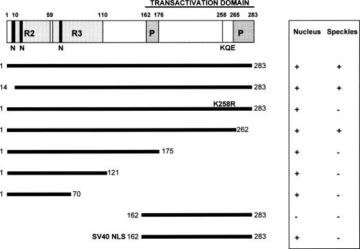
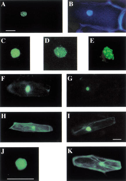
The photoreceptor phytochrome (phy) A has a well-defined role in regulating gene expression in response to specific light signals. Here, we describe a new Arabidopsis mutant, laf1 (long after far-red light 1) that has an elongated hypocotyl specifically under far-red light. Gene expression studies showed that laf1 has reduced responsiveness to continuous far-red light but retains wild-type responses to other light wavelengths. As far-red light is only perceived by phyA, our results suggest that LAF1 is specifically involved in phyA signal transduction. Further analyses revealed that laf1 is affected in a subset of phyA-dependent responses and the phenotype is more severe at low far-red fluence rates. LAF1 encodes a nuclear protein with strong homology with the R2R3-MYB family of DNA-binding proteins. Experiments using yeast cells identified a transactivation domain in the C-terminal portion of the protein. LAF1 is constitutively targeted to the nucleus by signals in its N-terminal portion, and the full-length protein accumulates in distinct nuclear speckles. This accumulation in speckles is abolished by a point mutation in a lysine residue (K258R), which might serve as a modification site by a small ubiquitin-like protein (SUMO).


Figures

Figure 1
Figure 1
laf1 seedlings are specifically impaired in FR-induced de-etiolation. (A) Hypocotyl lengths of laf1 compared with WT (Ler), phyA, and phyB under white (W; 15 μmole/m2 sec), far-red light (FR; 3 μmole/m2 sec), red light (R; 35 μmole/m2 sec) conditions and darkness (D). Error bars show standard deviations. (B) Phenotypes of laf1. Seedlings of WT (Ler), laf1, phyA, and phyB were grown for 4 d either in complete darkness under FR (3 μmole/m2 sec) or under R light (35 μmole/m2 sec). Bar, 10 mm.
Figure 2
Figure 2
LAF1 encodes a protein with homology with R2R3–MYB proteins. (A) A schematic representation of the genomic organization of laf1. The exon (black)/ intron (white) structure and the insertion site of the Ds element (506 bp from the ATG start codon) are shown. (B) Sequence comparison of LAF1 with other members of the R2R3–MYB protein family in Arabidopsis: AtMYB 19 (MYB19; GenBank accession no. Z95745), WEREWOLF (WER; Lee and Schiefelbein 1999), and Production of Anthocyanin Pigment 1 (PAP1; Borevitz et al. 2000). Conserved amino acid residues are highlighted in black. (Arrows) MYB domains (R2 and R3) ; (asterisks) conserved tryptophan residues within these domains. (C) Hypocotyl lengths of laf1 complemented with the 35S–LAF1 cDNA (c-1, c-2) or the 35S–LAF1 genomic transgene (g-1, g-2) and transgenic WT lines with the LAF1 antisense construct (as-1, as-2) or the 35S–LAF1 cDNA overexpressed (oe-1, oe-2). In comparison, the hypocotyl lengths of WT (Ler), phyA, and laf1 are shown. Light conditions are W (15 μmole/m2 sec), R (5 μmole/m2 sec), FR light at low fluence (1.5 μmole/m2 sec), and FR light at high fluence (6 μmole/m2 sec). Error bars show standard deviations.
Figure 3
Figure 3
Expression of CAB, CHS , and PET E in laf1. Four-day-old seedlings were grown either in the dark under different FR light fluencies (1.5, 3, and 6 μmole/m2 sec) or in W light for 18 h. Each lane contained 10 μg of total RNA. CAB, CHS, and PET E were used as probes. (A) Representative Northern blots. The hybridization with the 18S rDNA probe was used as a loading control. (B) Quantitative expression levels of CAB, CHS, and PET E in WT (Ler), laf1, and phyA. Northern blots were quantified with a PhosphoImager, and the expression levels were normalized with respect to the 18S rRNA level.
Figure 4
Figure 4
The laf1 mutant is specifically disrupted in phyA-dependent gene regulation. Four-day-old seedlings (Ler, phyA, phyB, and laf1) were grown in darkness and then, where indicated, transferred to R (35 μmole/m2 sec) or FR light (3 μmole/m2 sec) for 2 or 18 h. Each lane contained 10 μg of total RNA. CAB, CHS, and XTR7 were used as probes. The 18S rRNA band was used as a loading control.
Figure 5
Figure 5
LAF1 acts as a transcriptional activator. (A) Transactivation analysis of LAF1 in yeast. Different LAF1 cDNA fragments were fused to sequences encoding the Gal4 DNA-binding domain in the yeast vector pGBT8 and transformed into yeast strain HF7c. Transformants were plated onto SD plates with histidine and incubated for 3 d. Three different colonies were transferred to SD plates with histidine (SD + HIS), without histidine (SD) and without histidine plus 30 mM 3-AT (SD + 3-AT), and cell growth ability was analyzed after incubation for 4 d. (aa) Amino acid. (B) β-Galactosidase liquid culture assay from yeast carrying a pGBT8 vector either empty or with different LAF1 cDNA fragments by using ONPG as a substrate. The obtained β-galactosidase activity obtained from each construct was depicted relative to the basal levels obtained from the pGBT8 vector (=1). The data represent average values from three independent experiments in which several colonies were used to initiate the cultures.
Figure 6
Figure 6
Structure of LAF1–GFP fusion proteins and their respective localization to the nucleus and to nuclear speckles. A schematic diagram of the structure of the LAF1 protein. The MYB domain (R2, R3), the PEST domains (P), the nuclear localization signals (N), the putative sumoylation site (KQE), and the transactivation domain are indicated at the top (numbers indicate amino acid residues). The LAF1 fragments that were fused to the N terminus of GFP are illustrated by bars. SV40-NLS indicates a fusion with the SV40 nuclear localization signal. For analysis of the localization of fusion proteins at least 50 transformed cells were examined in at least three independent transient expression assays by using onion epidermal cells. The table on the right shows the localization of the various GFP fusion proteins (+) positive; (−) negative.
Figure 7
Figure 7
Representative images of subcellular location of LAF1–GFP fusions in transiently transformed onion epidermal cells. All experiments, except (D) were assayed 6–12 h after bombardment. (A) LAF1/1–283–GFP. (B) The same cell as in (A) stained with DAPI to show the location of the nucleus. (C) LAF1/1–283 3–6 h after bombardment. (D,E) LAF1/1–283 6–12 h after bombardment. (F) LAF1/1–70, (G) LAF1/1–175, (H) LAF1/162–283, (I) SV40-LAF1/162–283, (J) LAF1/1–283 K258R, (K) A control cell expressing GFP alone. Bars in A, I, and J, 50 μm.

References

    1. Altschul SF, Gish W, Miller W, Myers EW, Lipman DJ. Basic local alignment search tool. J Mol Biol. 1990;215:403–410. - PubMed
    1. Barnes SA, Nishizawa NK, Quaggio RB, Whitelam GC, Chua NH. Far-red light blocks greening of Arabidopsis seedlings via a phytochrome A-mediated change in plastid development. Plant Cell. 1996a;8:601–615. - PMC - PubMed
    1. Barnes SA, Quaggio RB, Whitelam GC, Chua NH. fhy1 defines a branch point in phytochrome A signal transduction pathways for gene expression. Plant J. 1996b;10:1155–1161. - PubMed
    1. Bolle C, Koncz C, Chua NH. PAT1, a new member of the GRAS family, is involved in phytochrome A signal transduction. Genes & Dev. 2000;14:1269–1278. - PMC - PubMed
    1. Borevitz JO, Xia Y, Blount J, Dixon RA, Lamb C. Activation tagging identifies a conserved MYB regulator of phenylpropanoid biosynthesis. Plant Cell. 2000;12:2383–2394. - PMC - PubMed

Publication types

MeSH terms