. 2001 Oct 1;155(1):123-31.
doi: 10.1083/jcb.200105110.

A nitric oxide synthase transgene ameliorates muscular dystrophy in mdx mice

Affiliations

A nitric oxide synthase transgene ameliorates muscular dystrophy in mdx mice

M Wehling et al. J Cell Biol. .

Abstract

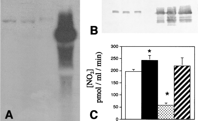
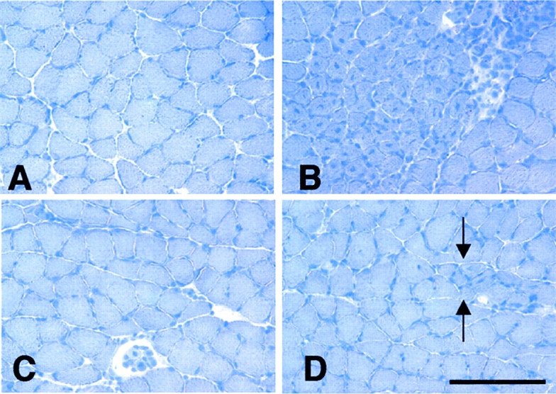
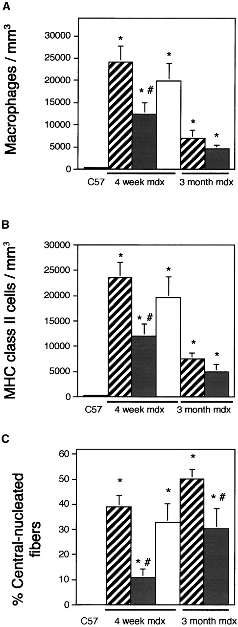
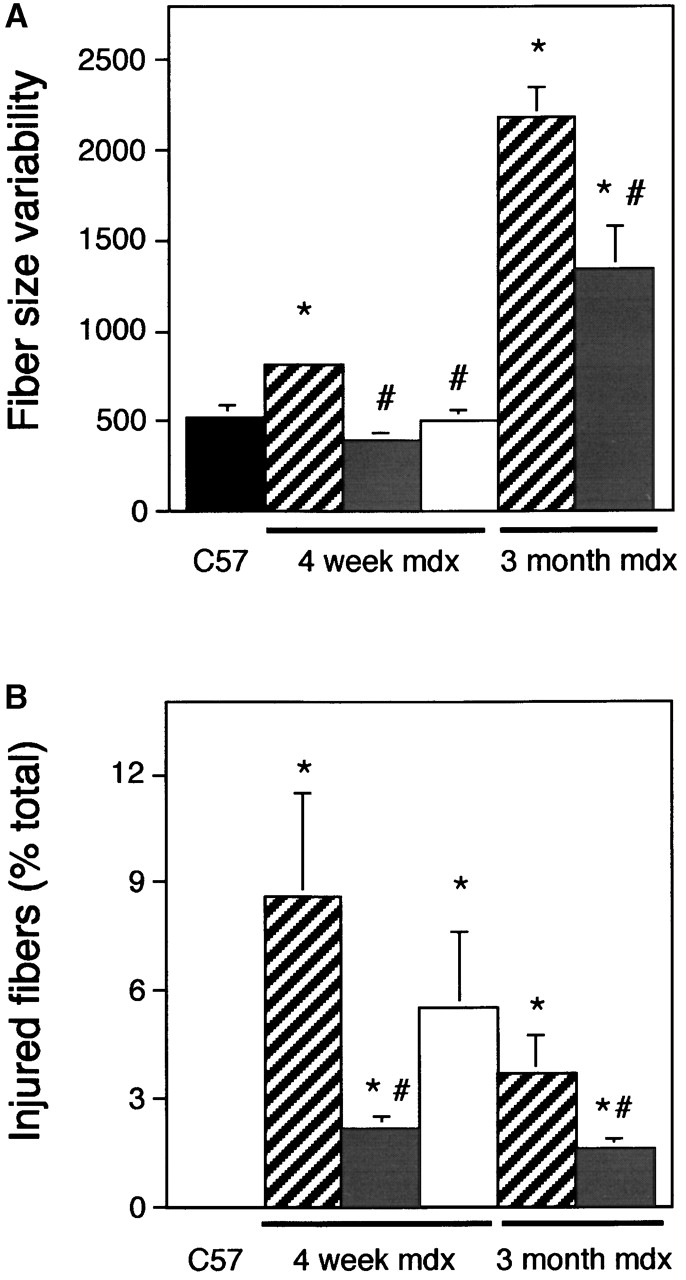
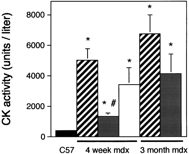
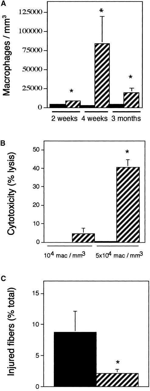
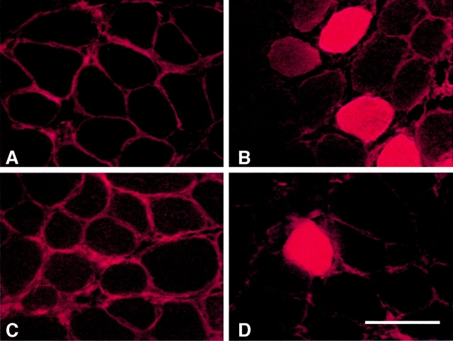
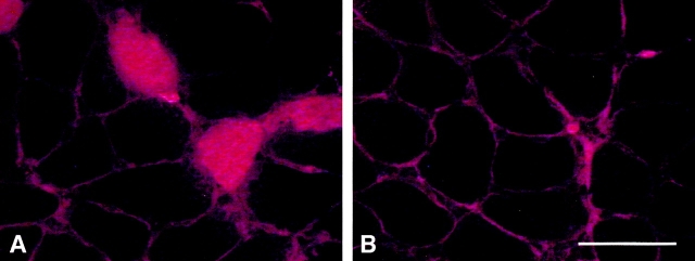
Dystrophin-deficient muscles experience large reductions in expression of nitric oxide synthase (NOS), which suggests that NO deficiency may influence the dystrophic pathology. Because NO can function as an antiinflammatory and cytoprotective molecule, we propose that the loss of NOS from dystrophic muscle exacerbates muscle inflammation and fiber damage by inflammatory cells. Analysis of transgenic mdx mice that were null mutants for dystrophin, but expressed normal levels of NO in muscle, showed that the normalization of NO production caused large reductions in macrophage concentrations in the mdx muscle. Expression of the NOS transgene in mdx muscle also prevented the majority of muscle membrane injury that is detectable in vivo, and resulted in large decreases in serum creatine kinase concentrations. Furthermore, our data show that mdx muscle macrophages are cytolytic at concentrations that occur in dystrophic, NOS-deficient muscle, but are not cytolytic at concentrations that occur in dystrophic mice that express the NOS transgene in muscle. Finally, our data show that antibody depletions of macrophages from mdx mice cause significant reductions in muscle membrane injury. Together, these findings indicate that macrophages promote injury of dystrophin-deficient muscle, and the loss of normal levels of NO production by dystrophic muscle exacerbates inflammation and membrane injury in muscular dystrophy.


Figures

Figure 1.
Figure 1.
Northern analysis of NOS Tg muscles. (A) Lanes 1–3 contained total RNA isolated from muscles that did not express the NOS transgene. Lane 4 was loaded with an equal mass of total RNA that was obtained from muscles of a NOS Tg mouse. The blot was probed with cDNA for full-length nNOS. The mass of the wild-type nNOS is greater than the transgenic nNOS, because the wild-type encodes an additional 34 amino acid nNOSμ peptide. (B) Western analysis of NOS Tg muscles. Muscle samples from littermates of NOS Tg mice that did not express the NOS transgene (three left lanes) and from NOS Tg mice (three right lanes). Densitometric analysis of the relative quantities of nNOS shows an ∼50-fold increase in nNOS in the NOS/Tg muscles. (C) NO release from control, NOS Tg, mdx, and NOS Tg/mdx. NO release from solei was measured for muscles obtained from C57 (white bar; n = 10), NOS Tg (black bar; n = 10), mdx (stippled bar; n = 8), and NOS Tg/mdx mice (striped; n = 8). Asterisk, differs from C57 at P < 0.05. Error bars indicate standard errors of the means in all figures.
Figure 2.
Figure 2.
Sections of 4-wk-old soleus muscles stained by hematoxylin. (A) C57 muscle showing fibers of uniform diameter, no central nucleation, and no clusters of inflammatory cells between adjacent fibers. (B) Mdx muscle showing a typical focus of muscle pathology characterized by fiber populations of variable diameters and central nucleation. Dark staining nuclei of inflammatory cells appear between adjacent fibers. (C) NOS Tg/mdx muscle showing typical histology, where fiber diameter is more uniform than age-matched mdx muscle, and there is little inflammation or central nucleation. (D) NOS Tg/mdx muscle showing an example of the relatively small lesions that appear in NOS Tg/mdx muscle (between arrows) where there are small clusters of small-diameter, central-nucleated fibers. All micrographs are at the same magnification. Bar, 250 μm.
Figure 3.
Figure 3.
Expression of the NOS transgene reduces mdx muscle inflammation and regeneration. The concentrations of macrophages (A), MHC class II+ cells (B), and regenerative fibers (C) were measured in quadriceps from 4-wk-old C57 (black bars; n = 10), 4-wk-old mdx (striped bars; n = 10), 4-wk-old NOS Tg/mdx (gray bars; n = 10), 4-wk-old, L-NAME–treated NOS Tg/mdx muscles (white bars; n = 6), 3-mo-old mdx (striped bars; n = 13), and 3-mo-old, NOS Tg/mdx (grey bars; n = 5). Asterisk, differs from C57 (P < 0.05); pound sign, differs from age-matched mdx (P < 0.05).
Figure 4.
Figure 4.
Expression of the NOS transgene reduces muscle fiber variability in size (A) and muscle fiber injury (B). Histogram shows variability in fiber diameter of 4-wk-old C57 (black bars; n = 10), 4-wk-old mdx (striped bars; n = 10), 4-wk-old NOS Tg/mdx (gray bars; n = 10), 4-wk-old, L-NAME–treated NOS Tg/mdx muscles (white bars; n = 6), 3-mo-old mdx (striped bars; n = 13), and 3-mo-old NOS Tg/mdx (grey bars; n = 5). (A) The data represent the mean of the standard deviation of fibers. (B) The data are the percentage of total fibers that contain procion red. Asterisk, differs from C57 (P < 0.05); pound sign, differs from age-matched mdx (P < 0.05).
Figure 6.
Figure 6.
Expression of the NOS transgene reduces the concentration of serum CK in mdx mice. 4-wk-old C57 (black bars; n = 5), 4-wk-old mdx (striped bars; n = 12), 4-wk-old NOS Tg/mdx (gray bars; n = 5), 4-wk-old, L-NAME–treated NOS Tg/mdx muscles (white bars; n = 6), 3-mo-old, mdx (striped bars; n = 13), and 3-mo-old NOS Tg/mdx (grey bars; n = 5). Asterisk, differs from C57 (P < 0.05); pound sign, differs from age-matched mdx (P < 0.05).
Figure 7.
Figure 7.
Macrophage concentration in mdx solei. (A) C57 (solid bars). Mdx (striped bars). Asterisk, differs from age-matched controls (P < 0.05). Pound sign, differs from all other data sets (P < 0.05). n = 14 for each data set. (B) Mdx muscle macrophages kill muscle cells in vitro. Values show the percentage lysis of C2C12 myotubes by macrophages isolated from 4-wk-old mdx muscles. Solid bars, nonactivated macrophages. Striped bars, PMA-activated macrophages. Asterisk, differs from C57 at P < 0.05. (C) Macrophage depletions reduce muscle membrane lesions. Data are the percentage of total muscle fibers in cross-sections of soleus muscle that contain intracellular procion red. Approximately 9% of the total fibers were injured and contained procion red in solei of mdx mice receiving intraperitoneal injections of PBS (black bar). Approximately 2% of the fibers of the fibers of mdx mice receiving intraperitoneal injections of anti-F4/80 showed intracellular procion red (striped bar). Asterisk, differs from PBS at P < 0.05.
Figure 5.
Figure 5.
Expression of the NOS transgene reduces muscle membrane lesions. Solei of C57 mice showed procion red almost exclusively located extracellularly (A), indicating infrequent occurrence of membrane lesions. Mdx muscles (B) showed that ∼8% of the fibers were injured and contained procion red. NOS Tg/mdx muscles (C and D) showed a frequency of procion red–containing injured fibers that was reduced to levels nearing C57 controls. Bar, 100 μm.
Figure 8.
Figure 8.
Macrophage depletions reduce mdx muscle fiber injury. Soleus muscles from 4-wk-old mdx mice showed a frequent occurrence of fibers with intracellular procion red, indicating the presence of membrane lesions (A). Depletion of circulating macrophages by anti-F4/80 injections decreased the occurrence of membrane lesions (B). Bar, 100 μm.

Similar articles

Cited by

References

    1. Abu-Soud, H.M., and S.L. Hazen. 2000. Nitric oxide modulates the catalytic activity of myeloperoxidase. J. Biol. Chem. 275:5425–5430. - PubMed
    1. Albina, J.E., S. Cui, R.B. Mateo, and J.S. Reichner. 1993. Nitric oxide-mediated apoptosis in murine peritoneal macrophages. J. Immunol. 150:5080–5085. - PubMed
    1. Amalfitano, A., and J.S. Chamberlain. 1996. The mdx-amplification-resistant mutation system assay, a simple and rapid polymerase chain reaction-based detection of the allele. Muscle Nerve. 19:1549–1553. - PubMed
    1. Anderson, J.E. 2000. A role for nitric oxide in muscle repair: nitric oxide-mediated activation of muscle satellite cells. Mol. Biol. Cell. 11:1859–1874. - PMC - PubMed
    1. Arahata, K., and A.G. Engel. 1984. Monoclonal antibody analysis of mononuclear cells in myopathies. I: Quantitation of subsets according to diagnosis and sites of accumulation and demonstration and counts of muscle fibers invaded by T cells. Ann. Neurol. 16:193–208. - PubMed

Publication types

MeSH terms