

Crystal structure of the TRANCE/RANKL cytokine reveals determinants of receptor-ligand specificity
- PMID: 11581298
- PMCID: PMC200957
- DOI: 10.1172/JCI13890
Crystal structure of the TRANCE/RANKL cytokine reveals determinants of receptor-ligand specificity
Abstract
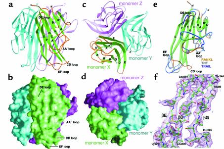
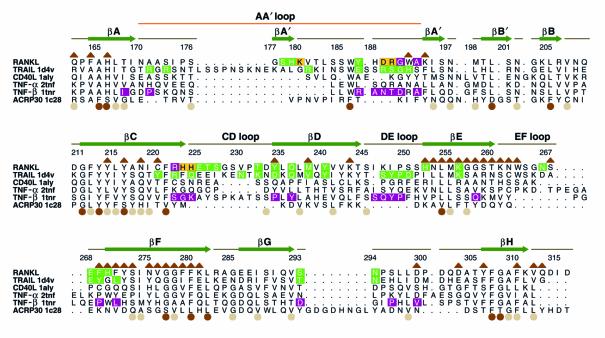
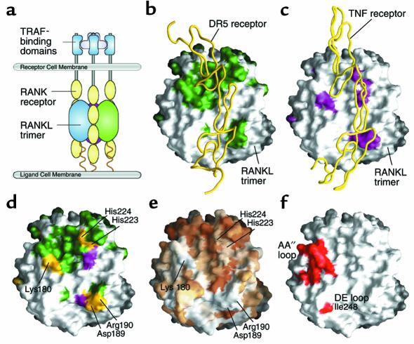
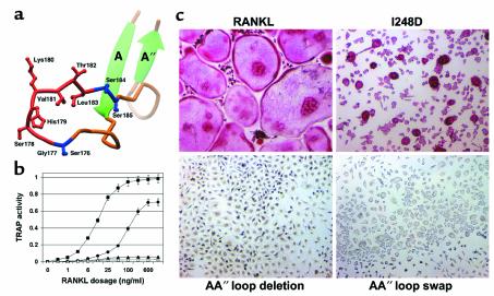
RANK, the receptor activator of NF-kappaB, and its ligand RANKL (initially termed TRANCE, also termed ODF and OPGL), are a TNF superfamily receptor-ligand pair that govern the development and function of osteoclasts, lymphoid tissue, and mammary epithelium. While TNF family cytokines share a common structural scaffold, individual receptor-ligand pairs associate with high specificity. Given the low level of amino acid conservation among members of the TNF superfamily, the means by which these molecules achieve specificity cannot be completely understood without knowledge of their three-dimensional structures. To determine the elements of RANKL that mediate RANK activation, we have crystallized the ectodomain of murine RANKL and solved its structure to a resolution of 2.6 A. RANKL self-associates as a homotrimer with four unique surface loops that distinguish it from other TNF family cytokines. Mutagenesis of selected residues in these loops significantly modulates RANK activation, as evidenced by in vitro osteoclastogenesis, thereby establishing their necessity in mediating the biological activities of RANKL. Such structural determinants of RANKL-RANK specificity may be of relevance in the pharmacologic design of compounds to ameliorate osteopenic disorders of bone.
Figures
Similar articles
-
A new member of tumor necrosis factor ligand family, ODF/OPGL/TRANCE/RANKL, regulates osteoclast differentiation and function.Biochem Biophys Res Commun. 1999 Mar 24;256(3):449-55. doi: 10.1006/bbrc.1999.0252. Biochem Biophys Res Commun. 1999. PMID: 10080918 Review.
-
Crystal structure of RANK ligand involved in bone metabolism.Vitam Horm. 2004;67:19-33. doi: 10.1016/S0083-6729(04)67002-6. Vitam Horm. 2004. PMID: 15110169 Review.
-
Crystal structure of the extracellular domain of mouse RANK ligand at 2.2-A resolution.J Biol Chem. 2002 Feb 22;277(8):6631-6. doi: 10.1074/jbc.M106525200. Epub 2001 Nov 30. J Biol Chem. 2002. PMID: 11733492
-
Expression profiles of receptor activator of nuclear factor kappaB ligand, receptor activator of nuclear factor kappaB, and osteoprotegerin messenger RNA in aged and ovariectomized rat bones.J Bone Miner Res. 2001 Aug;16(8):1416-25. doi: 10.1359/jbmr.2001.16.8.1416. J Bone Miner Res. 2001. PMID: 11499864
-
Promoter structure of mouse RANKL/TRANCE/OPGL/ODF gene.Biochim Biophys Acta. 1999 Apr 14;1445(1):134-41. doi: 10.1016/s0167-4781(99)00032-9. Biochim Biophys Acta. 1999. PMID: 10209265
Cited by
-
New perspectives on traumatic bone infections.Chin J Traumatol. 2020 Dec;23(6):314-318. doi: 10.1016/j.cjtee.2020.05.009. Epub 2020 Jun 2. Chin J Traumatol. 2020. PMID: 32847694 Free PMC article. Review.
-
Influence of Tumor Necrosis Factor α, Parathyroid Hormone, and Vitamin D3 on Modulation of the RANKL2 Isoform: A Pilot Study.Cartilage. 2012 Jan;3(1):100-3. doi: 10.1177/1947603511405837. Cartilage. 2012. PMID: 26069623 Free PMC article.
-
Bench to bedside: elucidation of the OPG-RANK-RANKL pathway and the development of denosumab.Nat Rev Drug Discov. 2012 May;11(5):401-19. doi: 10.1038/nrd3705. Nat Rev Drug Discov. 2012. PMID: 22543469 Review.
-
Structural insights into the binding of zoledronic acid with RANKL via computational simulations.Front Mol Biosci. 2022 Sep 19;9:992473. doi: 10.3389/fmolb.2022.992473. eCollection 2022. Front Mol Biosci. 2022. PMID: 36200071 Free PMC article.
-
RANKing c-Jun in osteoclast development.J Clin Invest. 2004 Aug;114(4):463-5. doi: 10.1172/JCI22644. J Clin Invest. 2004. PMID: 15314680 Free PMC article.
References
-
- Wong BR, et al. TRANCE is a novel ligand of the tumor necrosis factor receptor family that activates c-Jun N-terminal kinase in T cells. J Biol Chem. 1997;272:25190–25194. - PubMed
-
- Anderson DM, et al. A homologue of the TNF receptor and its ligand enhance T-cell growth and dendritic-cell function. Nature. 1997;390:175–179. - PubMed
-
- Lacey DL, et al. Osteoprotegerin ligand is a cytokine that regulates osteoclast differentiation and activation. Cell. 1998;93:165–176. - PubMed
Publication types
MeSH terms
Substances
Associated data
- Actions
Grants and funding
- AR-46852/AR/NIAMS NIH HHS/United States
- T32 AR007033/AR/NIAMS NIH HHS/United States
- R01 AR046523/AR/NIAMS NIH HHS/United States
- R37 AR046523/AR/NIAMS NIH HHS/United States
- R01 AR046852/AR/NIAMS NIH HHS/United States
- T32 GM007200/GM/NIGMS NIH HHS/United States
- R01 CA086803/CA/NCI NIH HHS/United States
- P50 GM062414/GM/NIGMS NIH HHS/United States
- AR-07033/AR/NIAMS NIH HHS/United States
- R01 AR032788/AR/NIAMS NIH HHS/United States
- GM-62414/GM/NIGMS NIH HHS/United States
- AR-32788/AR/NIAMS NIH HHS/United States
- CA-86803-01/CA/NCI NIH HHS/United States
- AR-46523/AR/NIAMS NIH HHS/United States
- Wellcome Trust/United Kingdom
- DE-05413/DE/NIDCR NIH HHS/United States
- GM-07200-25/GM/NIGMS NIH HHS/United States
LinkOut - more resources
Full Text Sources
Other Literature Sources
Molecular Biology Databases
