. 2001 Oct;108(7):991-9.
doi: 10.1172/JCI13345.

Transgenic expression of survivin in keratinocytes counteracts UVB-induced apoptosis and cooperates with loss of p53

Affiliations

Transgenic expression of survivin in keratinocytes counteracts UVB-induced apoptosis and cooperates with loss of p53

D Grossman et al. J Clin Invest. 2001 Oct.

Abstract

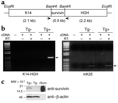
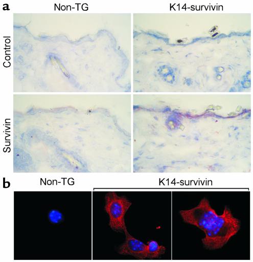
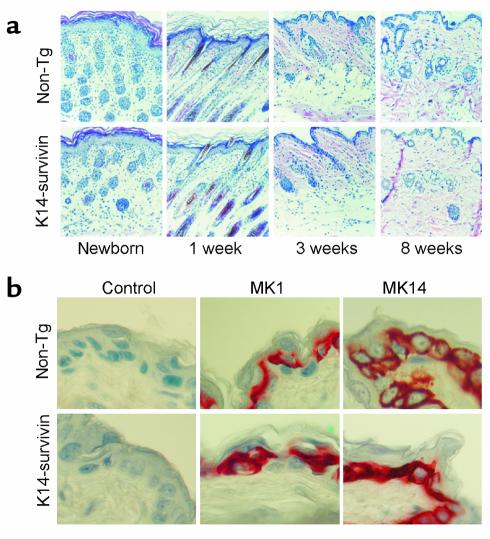
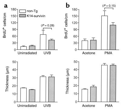
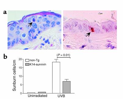
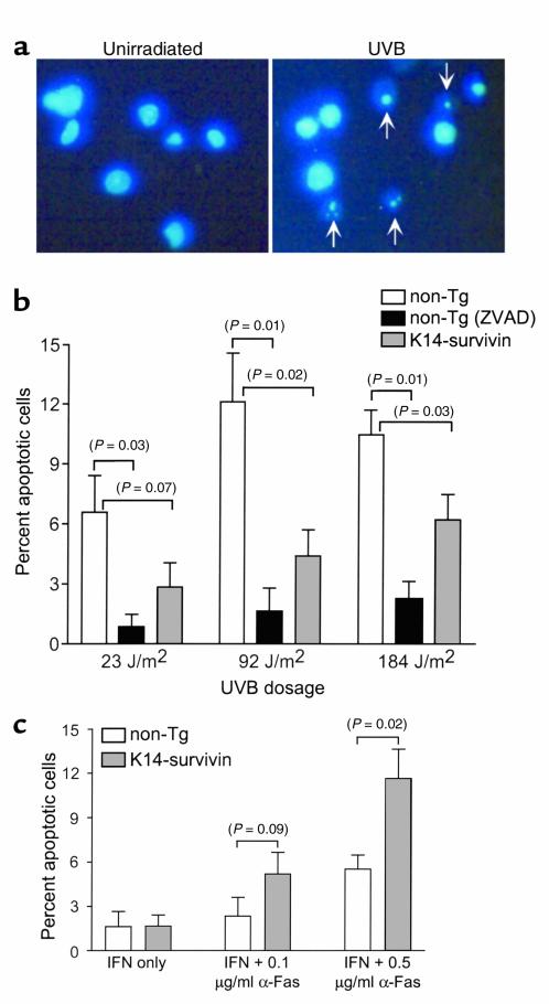
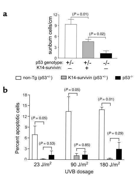
The inhibitor of apoptosis protein survivin has been implicated in both cell cycle control and apoptosis resistance. To discriminate between these different roles, we used transgenic expression of survivin in the skin as a model for cell proliferation, differentiation, and apoptosis. Transgenic mice expressing survivin under the control of a keratin-14 promoter developed normally, without histologic abnormalities of the skin or hair, epidermal hyperplasia, or developmental abnormalities of basal or suprabasal epidermis. Keratinocyte proliferation assessed under basal conditions, or after ultraviolet-B (UVB) irradiation, or phorbol ester stimulation was unchanged in survivin transgenic mice. In contrast, survivin expression inhibited UVB-induced apoptosis in vitro and in vivo (i.e., sunburn cell formation), whereas it did not affect Fas-induced cell death. When crossed with p53 knockout mice, transgenic expression of survivin in a p53(+/-) background substituted for the loss of a second p53 allele and further inhibited UVB-induced apoptosis. These data provide the first in vivo evidence that survivin inhibits apoptosis and suggest that this pathway may oppose the elimination of cancerous cells by p53.


Figures

Figure 1
Figure 1
Construction and characterization of K14-survivin transgenic mice. (a) The 0.5-kb mouse survivin cDNA, including the stop codon, was cloned into a BamHI site flanked by 2.1 kb of K14 promoter sequences and 2.2 kb of HGH and polyadenylation sequences, and the expression cassette was released from the vector by EcoRI digest. Arrows indicate the approximate location of PCR primers used for genotyping and RT-PCR. (b) RNA isolated from epidermal cells of K14-survivin transgenic (Tg+) and nontransgenic (Tg–) mice was incubated in the presence or absence of reverse transcriptase (RT) and amplified by PCR with primers complementary to K14 and HGH (left) or mouse keratin 2E (right). Molecular-weight (MW) markers are in the far left lanes, and arrows designate predicted products. (c) Lysates (50 μg) prepared from epidermal cells of K14-survivin (Tg+) and nontransgenic (Tg–) mice or aliquots (5 ng) of recombinant survivin (rSurv) were electrophoresed, transferred to nylon membranes, and blotted with Ab’s to survivin or β-actin, as indicated. Molecular-weight markers are shown on the left.
Figure 2
Figure 2
Immunohistochemical and subcellular localization of transgenic survivin expression. (a) Five-micrometer tissue sections were cut from fresh-frozen skin of K14-survivin transgenic mice (K14-survivin) or control nontransgenic (non-TG) littermates, fixed in ice-cold acetone, and analyzed by immunohistochemistry with Ab against survivin or a control Ab to EGFP. Binding of the primary Ab’s was visualized with a goat anti-rabbit polyclonal Ab using Vectastain Elite ABC and AEC peroxidase substrate kits. (b) Subcellular localization of transgenic survivin. Keratinocytes isolated from K14-survivin transgenic mice (K14-survivin) or nontransgenic littermates (non-TG) were adhered to glass coverslips, fixed in methanol-acetone, and incubated with an Ab to survivin, followed by Texas red–conjugated goat anti-rabbit Ab. Nuclei were stained with Hoechst 33342. Image merging analysis is shown.
Figure 3
Figure 3
Histologic analysis of epidermis in K14-survivin animals. (a) K14-survivin transgenic mice (K14-survivin) or nontransgenic littermates (non-TG) were genotyped at birth, sacrificed at the indicated time intervals, and dorsal skin was excised, processed, and stained with hematoxylin/eosin. (b) Skin sections isolated from K14-survivin or non-TG control littermates were analyzed for reactivity with Ab’s to mouse cytokeratin-1 (MK1) or -14 (MK14), or nonimmune rabbit IgG (control) by immunohistochemistry.
Figure 4
Figure 4
Effect of transgenic survivin expression on keratinocyte proliferation and skin hyperplasia in vivo. (a) Nontransgenic littermates (white bars) and K14-survivin mice (gray bars) were irradiated with 600 J/m2 UVB and 72 hours later injected with BrdU. After 2 hours, unshaven (unirradiated) and shaved (UVB-treated) dorsal skin was excised and processed. BrdU-labeled cells were detected by immunohistochemistry, and epidermal thickness was measured. Data are expressed as number of BrdU-positive cells per linear centimeter of interfollicular epidermis (top) and as epidermal thickness (bottom). Error bars indicate SEM for five mice per group. P value for nontransgenic and K14-survivin mice is noted above bars in parentheses. (b) Nontransgenic littermates (white bars) and K14-survivin mice (gray bars) were shaved and subjected to topical applications of acetone or acetone containing PMA every 3 days. Forty-eight hours after the fourth application, mice were injected with BrdU, and BrdU-labeled cells and epidermal thickness were assessed as above. Error bars indicate SEM for seven mice per group. P value for nontransgenic and K14-survivin mice is noted above error bars in parentheses.
Figure 5
Figure 5
Effect of transgenic survivin on keratinocyte apoptosis in vivo. (a) Sections of UVB-irradiated (600 J/m2) nontransgenic mouse skin were stained with hematoxylin/eosin (left) or for internucleosomal DNA fragmentation by TUNEL (right). Arrows indicate an apoptotic keratinocyte (sunburn cell). (b) Quantitation of UVB-induced apoptosis in vivo. K14-survivin transgenic mice (gray bars) or nontransgenic littermates (white bars) were irradiated with UVB, and 24 hours later exposed skin was excised and sunburn cells were identified and counted by light microscopy and hematoxylin/eosin staining. Data are expressed as number of sunburn cells per linear centimeter of skin analyzed. Error bars indicate SEM of 2 and nine mice per group of unirradiated and irradiated mice, respectively. P value for nontransgenic and K14-survivin mice is noted in parentheses.
Figure 6
Figure 6
Characterization of survivin-dependent cytoprotection in transgenic keratinocytes. (a) Isolated keratinocytes were untreated or irradiated with UVB (90 J/m2) in vitro, and 24 hours later fixed, stained with DAPI, and analyzed by fluorescence microscopy. Arrows indicate apoptotic keratinocytes with condensed and fragmented nuclei. (b) Keratinocytes isolated from nontransgenic littermates (white bars) or K14-survivin transgenic mice (gray bars) were exposed to the indicated UVB doses in vitro, then fixed and stained with DAPI. Keratinocytes from nontransgenic littermates were also separately incubated in the presence of the pan-caspase inhibitor, ZVAD-fmk (black bars). Data are expressed as percentage of apoptotic cells based on counting approximately 150 cells. (c) Effect of transgenic survivin on IFN-γ/anti-Fas–induced apoptosis. Keratinocytes isolated from nontransgenic littermates (white bars) or K14-survivin mice (gray bars) were cultured with 1,000 U/ml IFN-γ alone or in combination with 0.1 or 0.5 μg/ml anti-Fas. After 24 hours, cells were fixed, stained with DAPI, and analyzed for nuclear morphology of apoptosis. Data are expressed as percentage of apoptotic cells based on counting approximately 120–150 cells. For each condition, the background level of apoptosis (ranging from 5 to 10%) in unirradiated cells has been subtracted. Error bars indicate SEM of four independent experiments. P values are noted in parentheses.
Figure 7
Figure 7
Survivin cooperates with loss of p53 in inhibiting UVB-induced apoptosis. (a) Nontransgenic (p53+/–; white bars), K14-survivin (p53+/–; gray bars), and p53 knockout (p53–/–; black bars) mice were irradiated with 600 J/m2 UVB, and sunburn cells were quantitated by hematoxylin/eosin staining. Data are expressed as number of sunburn cells per linear centimeter of skin analyzed. Error bars indicate SEM of 7, 11, and 3 mice per group of nontransgenic p53+/–, K14-survivin p53+/–, and p53–/– mice, respectively. (b) Keratinocytes isolated from nontransgenic p53+/– (white bars), K14-survivin p53+/– (gray bars), and p53–/– (black bars) mice were irradiated with UVB in vitro at the indicated doses. After 24 hours, cells were fixed, stained with DAPI, and analyzed for nuclear morphology of apoptosis. Error bars indicate SEM of three independent experiments. P values are noted in parentheses.

Comment in

  • The Survivin saga goes in vivo.
    Reed JC. Reed JC. J Clin Invest. 2001 Oct;108(7):965-9. doi: 10.1172/JCI14123. J Clin Invest. 2001. PMID: 11581297 Free PMC article. Review. No abstract available.

References

    1. Fuchs E. Keratins and the skin. Annu Rev Cell Dev Biol. 1995;11:123–153. - PubMed
    1. Taylor G, Lehrer MS, Jensen PJ, Sun TT, Lavker RM. Involvement of follicular stem cells in forming not only the follicle but also the epidermis. Cell. 2000;102:451–461. - PubMed
    1. Ishida-Yamamoto A, et al. Programmed cell death in normal epidermis and loricrin keratoderma. Multiple functions of profilaggrin in keratinization. J Investig Dermatol Symp Proc. 1999;4:145–149. - PubMed
    1. McCall CA, Cohen JJ. Programmed cell death in terminally differentiating keratinocytes: role of endogenous endonuclease. J Invest Dermatol. 1991;97:111–114. - PubMed
    1. Polakowska RR, Piacentini M, Bartlett R, Goldsmith LA, Haake AR. Apoptosis in human skin development: morphogenesis, periderm, and stem cells. Dev Dyn. 1994;199:176–188. - PubMed

Publication types

MeSH terms