. 2001 Oct;108(7):1001-13.
doi: 10.1172/JCI12864.

Inhibition of RXR and PPARgamma ameliorates diet-induced obesity and type 2 diabetes

Affiliations

Inhibition of RXR and PPARgamma ameliorates diet-induced obesity and type 2 diabetes

T Yamauchi et al. J Clin Invest. 2001 Oct.

Abstract

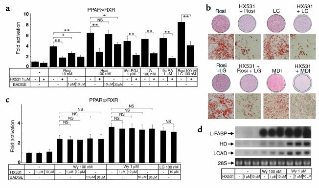
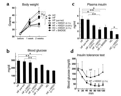
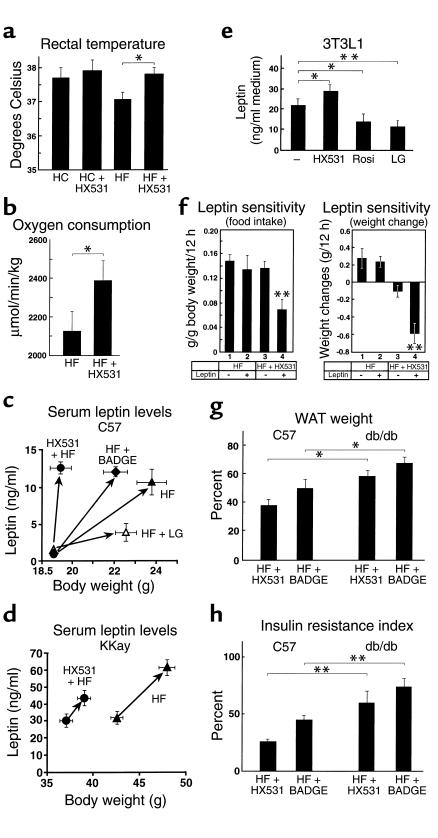
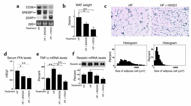
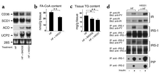
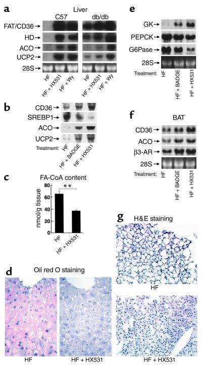
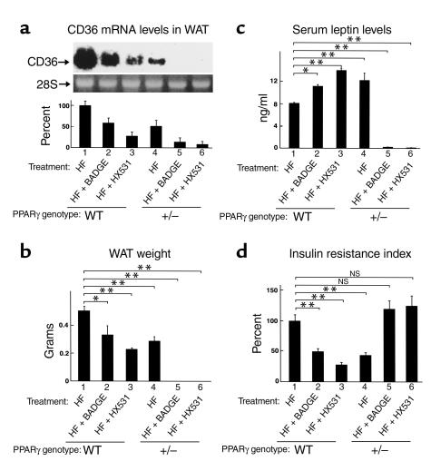
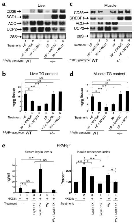
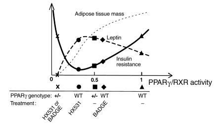
PPARgamma is a ligand-activated transcription factor and functions as a heterodimer with a retinoid X receptor (RXR). Supraphysiological activation of PPARgamma by thiazolidinediones can reduce insulin resistance and hyperglycemia in type 2 diabetes, but these drugs can also cause weight gain. Quite unexpectedly, a moderate reduction of PPARgamma activity observed in heterozygous PPARgamma-deficient mice or the Pro12Ala polymorphism in human PPARgamma, has been shown to prevent insulin resistance and obesity induced by a high-fat diet. In this study, we investigated whether functional antagonism toward PPARgamma/RXR could be used to treat obesity and type 2 diabetes. We show herein that an RXR antagonist and a PPARgamma antagonist decrease triglyceride (TG) content in white adipose tissue, skeletal muscle, and liver. These inhibitors potentiated leptin's effects and increased fatty acid combustion and energy dissipation, thereby ameliorating HF diet-induced obesity and insulin resistance. Paradoxically, treatment of heterozygous PPARgamma-deficient mice with an RXR antagonist or a PPARgamma antagonist depletes white adipose tissue and markedly decreases leptin levels and energy dissipation, which increases TG content in skeletal muscle and the liver, thereby leading to the re-emergence of insulin resistance. Our data suggested that appropriate functional antagonism of PPARgamma/RXR may be a logical approach to protection against obesity and related diseases such as type 2 diabetes.


Figures

Figure 1
Figure 1
The RXR antagonist HX531 serves as a functional PPARγ/RXR inhibitor (a and c). Transactivation analysis of PPARγ/RXR (a), PPARα/RXR (c). CV-1 cells were cotransfected with RXRα with an expression vector for PPARγ or PPARα. PPARγ/RXR or PPARα/RXR activity was assessed on a PPRE-tk LUC (24) as described previously (22, 23). CV-1 cells were treated with the indicated concentrations of rosiglitazone (Rosi), LG100268 (LG), 15-deoxy-Δ12,14 prostaglandin J2 (15d-PGJ2), Wy-14,643 (Wy), BADGE, and HX531. 9-cis-retinoic acid (b) 3T3L1 adipocyte differentiation assay. Oil red O staining for fat accumulation in cells at day 6 after induction. Cells were grown to confluence and then induced to differentiate by exposure to 100 nM rosiglitazone, 1 μM LG100268, or 10 μM HX531, or by conventional hormonal stimuli (MDI; a combination of 3-isobutyl-1-methylxanthine, dexamethasone, and insulin). (d) Amounts of the mRNAs of liver-type fatty acid–binding protein (L-FABP), HD, and long-chain acyl-CoA dehydrogenase (LCAD) in rat hepatoma FAO cells treated with the indicated concentrations of Wy-14,643 and HX531 for 24 hours. Each bar represents the mean ± SE (n = 5–10). *P < 0.05, **P < 0.01 with versus without HX531 or BADGE. NS, no significant difference.
Figure 2
Figure 2
Both the RXR antagonist HX531 and the PPARγ antagonist BADGE exert antiobesity and antidiabetic effects in proportion to their potencies as PPARγ/RXR inhibitors in vitro (ad). Body weight (a), fasting blood glucose (b), fasting plasma insulin (c), and insulin tolerance test (d) of KKAy mice untreated or treated with HX531 (+HX531) or BADGE (+BADGE) for 2 weeks while on the HF diet or the HC diet. HX531 or BADGE was given as an indicated percentage of food admixture. The same amounts of food were given to the pair-fed group as to mice treated with HX531, given as a 0.1% food admixture. Each bar represents the mean ± SE (n = 5–10). *P < 0.05, **P < 0.01 with versus without HX531 or BADGE.
Figure 3
Figure 3
Both the RXR antagonist HX531 and the PPARγ antagonist BADGE exert antiobesity and antidiabetic effects in part through leptin-dependent pathways (a and b). Rectal temperature (a) and oxygen consumption (b) of KKAy mice untreated or treated with HX531 (+HX531) for 2 weeks while on the HF diet or the HC diet. Serum leptin levels of C57 (c) or KKAy mice (d) untreated or treated with HX531, BADGE (+BADGE), or LG100268 (+LG) for 2 weeks while on the HF diet. The left sides of the arrows are before-treatment (c) and after 1-week treatment (d) levels. (e) Leptin protein levels in the medium of 3T3L1 adipocytes treated with 10 μM HX531, 1 μM LG100268, or 100 nM rosiglitazone (Rosi) for 24 hours. (f) Effects of intraperitoneal leptin administration in untreated C57 mice or C57 mice treated with HX531 for 10 days while on the HF diet. Groups of untreated mice or mice treated with HX531 received an intraperitoneal injection of either leptin (10 μg/g/d) (+) or isotonic sodium chloride solution (–). Food intake/12 h (left) and weight changes/12 h (right) were measured. (g and h) WAT weight (g) and insulin resistance index (7) (h) of C57 and db/db mice untreated or treated with HX531 or BADGE for 2 weeks while on the HF diet. The results are expressed as the percentage of the value of untreated mice on the HF diet (g and h). Each bar represents the mean ± SE (n = 5–10). *P < 0.05, **P < 0.01, C57 versus db/db or untreated versus treated with HX531, BADGE, Rosi, LG, or leptin.
Figure 4
Figure 4
Both the RXR antagonist HX531 and the PPARγ antagonist BADGE reduce expressions of the molecules involved in fatty acid influx and lipogenesis and increase expression of β3-AR in WAT. (a) Amounts of the mRNAs of FAT/CD36, SREBP1, and β3-AR in WAT (a) of KKAy mice. (b and c) WAT mass (b), histological analysis (top), and cell size distribution (bottom) (c) of epididymal WAT from KKAy mice. (df) Serum FFA levels (d), TNF-α mRNA levels (e), and resistin mRNA levels in WAT (f) of KKAy mice, untreated (HF), treated with HX531 (HF + HX531), or with BADGE (HF + BADGE) for 2 weeks while on the HF diet. HX531 or BADGE was given as a 0.1% or 3% food admixture, respectively. Each bar represents the mean ± SE (n = 5–10). *P < 0.05, **P < 0.01, HF versus HF + HX531 or HF + BADGE.
Figure 5
Figure 5
The RXR antagonist HX531 and the PPARγ antagonist BADGE both decrease molecules involved in fatty acid influx and lipogenesis and increase molecules involved in energy consumption in skeletal muscle. (a) Amounts of FAT/CD36, SCD1, ACO, and UCP2 in skeletal muscles (a) of KKAy mice. (bd) FA-CoA (b) and TG (c) content and expression and insulin-induced tyrosine phosphorylation of IR, IRS-1 and -2, and insulin-stimulated PI 3-kinase activity (d) in skeletal muscles of KKAy mice, untreated (HF) or treated with HX531 (HF + HX531) or with BADGE (HF + BADGE) for 2 weeks while on the HF diet. (b and c) Tissue homogenates were extracted, and their TG and FA-CoA content was determined as described previously (22, 26), with some modifications. (d) Mice were stimulated with or without 1 Ug–1 body weight of insulin for 2 minutes. Lysates were immunoprecipitated (IP) with the Ab’s indicated, followed by immunoblotting with the Ab’s indicated or kinase assay for PI. Labeled PI (PIP) was subjected to thin-layer chromatography and autoradiography as described previously (21). HX531 or BADGE was given as a 0.1% or 3% food admixture, respectively. Each bar represents the mean ± SE (n = 5–10). *P < 0.05, **P < 0.01, HF versus HF + HX531 or HF + BADGE.
Figure 6
Figure 6
The RXR antagonist HX531 and the PPARγ antagonist BADGE both increase molecules involved in fatty acid combustion and energy dissipation via leptin and PPARα pathways in the liver and BAT (a, b, e, f). Amounts of the mRNAs of FAT/CD36, SREBP1, HD (peroxisomal enoyl-CoA hydratase/3-hydroxyacyl-CoA dehydrogenase), ACO, UCP2, and β3-AR in livers from C57 and db/db mice (a), in livers from KKAy mice (b), in BAT from KKAy mice (f), and glucokinase, PEPCK, and G6Pase in livers from KKAy mice (e). (c, d, g) Hepatic FA-CoA content (c), hepatic TG content (oil red O staining) (d), size of brown adipocytes from KKAy mice (g), untreated (HF) or treated with HX531 (HF + HX531), Wy-14,643 (HF + Wy), or BADGE (HF + BADGE) for 2 weeks while on the HF diet. HX531, or Wy-14,643, or BADGE was given as a 0.1%, 0.01%, or 3% food admixture, respectively. Each bar represents the mean ± SE (n = 5–10). *P < 0.05, **P < 0.01, HF versus HF + HX531 or HF + BADGE.
Figure 7
Figure 7
Treatment of heterozygous PPARγ-deficient mice with an RXR antagonist or PPARγ antagonist results in re-emergence of hyperglycemia and insulin resistance associated with lipoatrophy. (a) Amounts of the mRNAs of FAT/CD36 in WAT. (bd) WAT mass (b), serum leptin levels (c), insulin resistance index (d), of wild-type (WT) and heterozygous PPARγ-deficient mice (+/–) untreated (–) or treated with HX531 or BADGE for 3 weeks (a) or for 4 weeks (b–d) while on the HF diet. HX531 or BADGE was given as a 0.1% or 3% food admixture, respectively. Each bar represents the mean ± SE (n = 5–10). *P < 0.05, **P < 0.01, untreated versus treated with HX531 or BADGE or compared with untreated wild-type mice. NS, no significant difference.
Figure 8
Figure 8
The combination of leptin deficiency and decreased effects of PPARα pathways results in insulin resistance in heterozygous PPARγ-deficient mice without WAT induced by treatment with an RXR antagonist or a PPARγ antagonist (a and c). Amounts of the mRNAs of FAT/CD36, SREBP1, SCD1, ACO, and UCP2 in the liver (a) and in skeletal muscle (c). (b and d) TG content of the livers (b) and skeletal muscles (d) of wild-type (WT) and heterozygous PPARγ-deficient mice (+/–) untreated (–) or treated with HX531 or BADGE for 4 weeks (ad) while on the HF diet. (e) Serum leptin levels (left) and insulin resistance index (right) of heterozygous PPARγ-deficient mice (PPARγ+/–) untreated (–) or treated with HX531 (+) for 6 weeks or PPARγ+/– treated with low doses of leptin (Lep1X), high doses of leptin (Lep10X), Wy-14,643 (Wy), or a combination for the last 12 days of a 6-week HX531 treatment while on the HF diet. HX531 or Wy-14,643 was given as a 0.1 or 0.01% food admixture, respectively. The results are expressed as the percentage of the value of untreated PPARγ+/– on the HF diet (right). HX531 or Wy-14,643 was given as a 0.1 or 0.01% food admixture, respectively. Each bar represents the mean ± SE (n = 5–10). *P < 0.05, **P < 0.01, untreated versus treated with HX531, Lep1X, Lep10X, or Wy, or compared with untreated wild-type mice. NS, no significant difference.
Figure 9
Figure 9
One possible model for the relationship between PPARγ/RXR activity and insulin sensitivity on the HF diet. The relationship between PPARγ/RXR activity and WAT mass may be linear. There may appear to be an optimal level of PPARγ/RXR activity for insulin sensitivity that is approximately 0.3–0.5 times normal. Increases in PPARγ/RXR activity as compared with the optimal range are associated with decreased serum leptin levels due to increased PPARγ/RXR-mediated suppression of leptin gene transcription, and decreases in PPARγ/RXR activity may be associated with decreased serum leptin levels due to depletion of WAT. Thus, these data raise the possibility that the relationship between PPARγ/RXR activity and leptin is an inverted U-shaped curve. The relationship between PPARγ/RXR activity and insulin resistance appears to exhibit such a U-shaped curve. Thus, impairment of the leptin pathway may closely parallel impairment of insulin sensitivity.

References

    1. Kersten S, Desvergne B, Wahli W. Roles of PPARs in health and disease. Nature. 2000;405:421–424. - PubMed
    1. Lowell BB. PPARgamma: an essential regulator of adipogenesis and modulator of fat cell function. Cell. 1999;99:239–242. - PubMed
    1. Spiegelman BM, Flier JS. Adipogenesis and obesity: rounding out the big picture. Cell. 1996;87:377–389. - PubMed
    1. Gonzalez FJ. Recent update on the PPAR alpha-null mouse. Biochimie. 1997;79:139–144. - PubMed
    1. Auwerx J. PPARgamma, the ultimate thrifty gene. Diabetologia. 1999;42:1033–1049. - PubMed

Publication types

MeSH terms