. 2001 Oct;108(7):1079-85.
doi: 10.1172/JCI12954.

Central melanocortin receptors regulate insulin action

Affiliations

Central melanocortin receptors regulate insulin action

S Obici et al. J Clin Invest. 2001 Oct.

Abstract

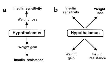
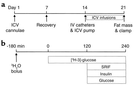
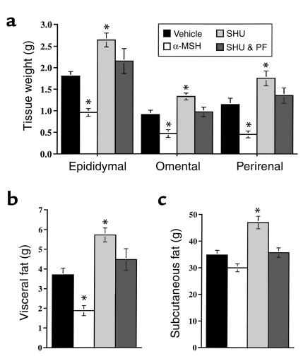
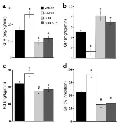
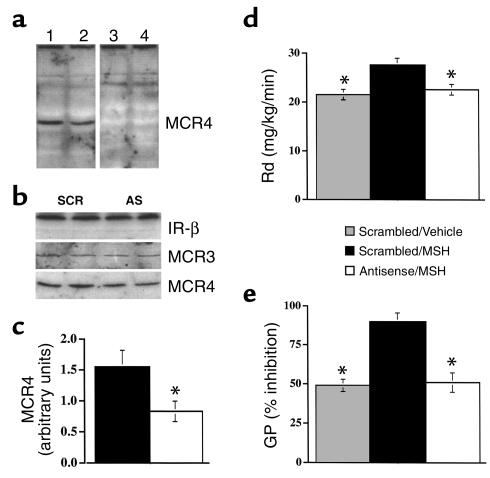
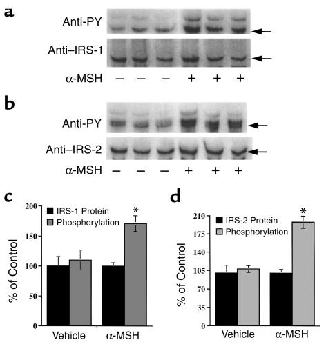
Energy balance and insulin action are tightly coregulated. Leptin regulates energy intake and expenditure partly by modulation of the melanocortin pathway in the hypothalamus. Here we demonstrate potent effects of the melanocortin pathway on insulin action and body distribution of adiposity. Conscious rats received week-long infusions of either a melanocortin receptor agonist, alpha-melanocyte-stimulating hormone (alpha-MSH), or antagonist, SHU9119, in the third cerebral ventricle while food intake was maintained constant in each group. alpha-MSH decreased intra-abdominal fat and markedly enhanced the actions of insulin on both glucose uptake and production, while SHU9119 exerted opposite effects. Our findings elucidate a neuroendocrine network that is likely to play a central role in the coupling of energy intake and insulin action.


Figures

Figure 1
Figure 1
Hypotheses on the regulation of insulin action by hypothalamic neural pathways. (a) Sequential model: neuronal pathways regulate feeding behavior and energy balance, which in turn modulate insulin action. (b) Parallel model: neuronal pathways concomitantly regulate weight homeostasis and insulin action. The two models are not mutually exclusive.
Figure 2
Figure 2
Schematic representation of the experimental design. (a) Surgical implantation of intracerebroventricular cannulae was performed on day 1 (∼3 weeks before the in vivo study). Full recovery of body weight and food intake was achieved by day 7. Surgical implantation of intravenous catheters and of osmotic minipumps (for intracerebroventricular [ICV] infusions) was performed on day 14. Intracerebroventricular infusions were continued for 7 days. Finally, on day 21, body composition and insulin action were estimated. (b) A bolus of tritiated water was given intravenously 3 hours before starting the tritiated glucose infusion. At t = 0 a primed-continuous infusion of labeled glucose was initiated and maintained for the remainder of the 4-hour study. Pancreatic-insulin clamp study was initiated at t = 120 minutes and lasted 120 minutes.
Figure 3
Figure 3
Effect of α-MSH and SHU9119 administration on subcutaneous and visceral fat. (a) Weight of epidydimal, omental, and perirenal fat depots in the four experimental groups. (b) Total visceral fat mass represents the sum of omental, epididymal, and perirenal fat depots. (c) Subcutaneous fat mass was estimated by subtracting the total visceral fat from the whole body fat mass. Of note, administration of α-MSH selectively decreased the size of all visceral fat depots compared with SHU-PF, despite similar food intake, weight changes, and subcutaneous fat mass. Values represent mean ± SE. *P < 0.01 vs. vehicle.
Figure 4
Figure 4
Role of the melanocortin pathway in the regulation of insulin action on peripheral glucose uptake and production. (a) During insulin clamp studies, the rate of glucose infusion was markedly increased by α-MSH and markedly decreased by SHU9119. (c) Insulin action on glucose uptake (Rd) was significantly enhanced by α-MSH and decreased by SHU9119. (b) During insulin clamp studies, the rate of glucose production (GP) was lower in rats treated with α-MSH and higher in rats receiving SHU9119, compared with vehicle. (d) The inhibition of GP in response to physiological hyperinsulinemia was markedly increased by α-MSH and markedly decreased by SHU9119. Values represent mean ± SE. *P < 0.01 vs. vehicle. GIR, glucose infusion rate.
Figure 5
Figure 5
The MCR4 mediates the effect of α-MSH on hepatic insulin action. (a) Western blot of MCR4 in hypothalamus of rats receiving intracerebroventricular infusions of either scrambled ODN (SCR; lanes 1 and 3) or MCR4 antisense (AS; lanes 2 and 4) for 7 days. Representative blots are displayed. The identity of the 43-kDa band was confirmed by peptide competition. Addition of a specific MCR4 peptide eliminated this band (lanes 3 and 4), but failed to alter a nonspecific band. (b) Intracerebroventricular administration of MCR4 antisense decreased hypothalamic MCR4 protein, while insulin receptor IR-β and MCR3 proteins were not affected. (c) Quantitation of hypothalamic Western blots: intracerebroventricular administration of MCR4 antisense led to a consistent, approximately 50% decrease in hypothalamic MCR4 protein. (d) During insulin clamp studies, the rate of glucose disappearance (Rd) was increased in rats treated with α-MSH with scrambled ODN; however, α-MSH failed to increase Rd in rats receiving MCR4 antisense. (e) During insulin clamp studies, the rate of glucose production (GP) was lower in rats treated with α-MSH with scrambled ODN; however, α-MSH failed to decrease GP in rats receiving MCR4 antisense. Values represent mean ± SE. *P < 0.01 vs. vehicle.
Figure 6
Figure 6
Effect of α-MSH on hepatic insulin signaling. Liver samples were immunoprecipitated with anti–IRS-1 and anti–IRS-2 Ab’s and then immunoblotted with anti-phosphotyrosine and anti–IRS1/2 Ab’s. IRS-1 and IRS-2 proteins were not affected by treatment with α-MSH. By contrast, tyrosine phosphorylation of both insulin receptor substrates was markedly increased following α-MSH treatment. Values represent mean ± SE. *P < 0.01 vs. vehicle.

Comment in

References

    1. McGarry JD. What if Minkowski had been ageusic? An alternative angle on diabetes. Science. 1992;258:766–770. - PubMed
    1. Petersen KF, et al. 13C/31P NMR studies on the mechanism of insulin resistance in obesity. Diabetes. 1998;47:381–386. - PubMed
    1. Zhang Y, et al. Positional cloning of the mouse obese gene and its human homologue. Nature. 1994;372:425–32. - PubMed
    1. Weigle DS, et al. Recombinant ob protein reduces feeding and body weight in the ob/ob mouse. J Clin Invest. 1995;96:2065–2070. - PMC - PubMed
    1. Campfield L, Smith FJ, Guisez Y, Devos R, Burn P. Recombinant mouse OB protein: evidence for a peripheral signal linking adiposity and central neural networks. Science. 1995;269:546–549. - PubMed

Publication types

MeSH terms