. 2001 Nov;75(21):10073-89.
doi: 10.1128/JVI.75.21.10073-10089.2001.

Macrophage tropism of human immunodeficiency virus type 1 isolates from brain and lymphoid tissues predicts neurotropism independent of coreceptor specificity

Affiliations

Macrophage tropism of human immunodeficiency virus type 1 isolates from brain and lymphoid tissues predicts neurotropism independent of coreceptor specificity

P R Gorry et al. J Virol. 2001 Nov.

Abstract

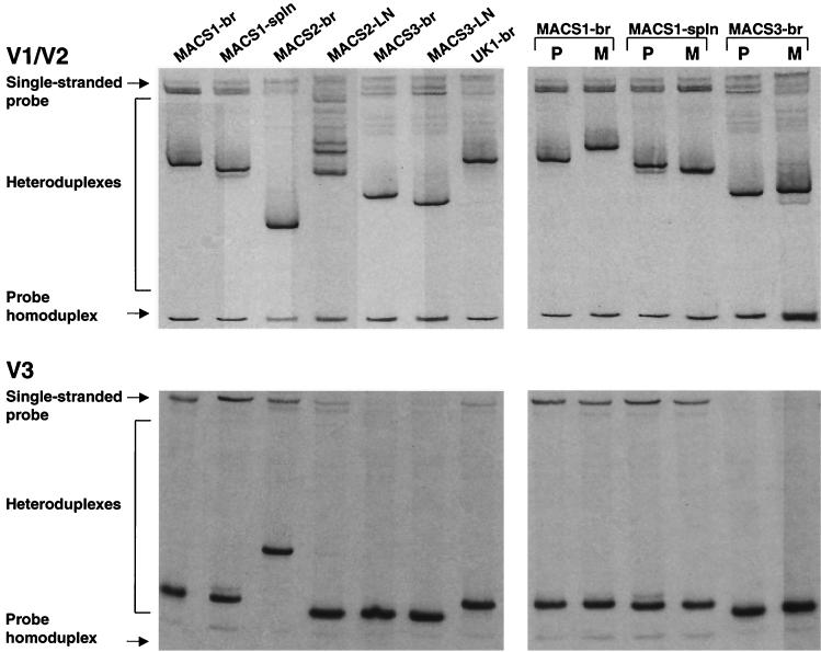
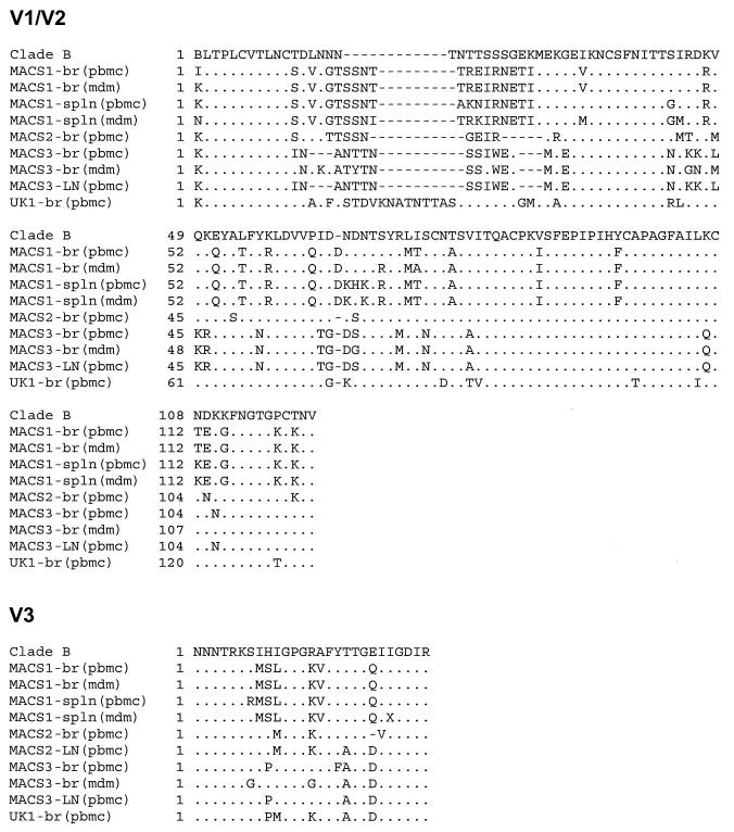
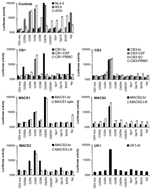
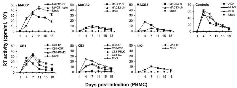
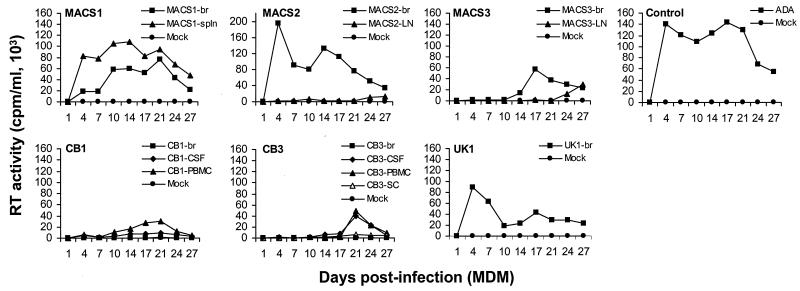
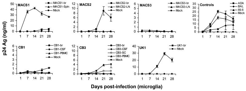
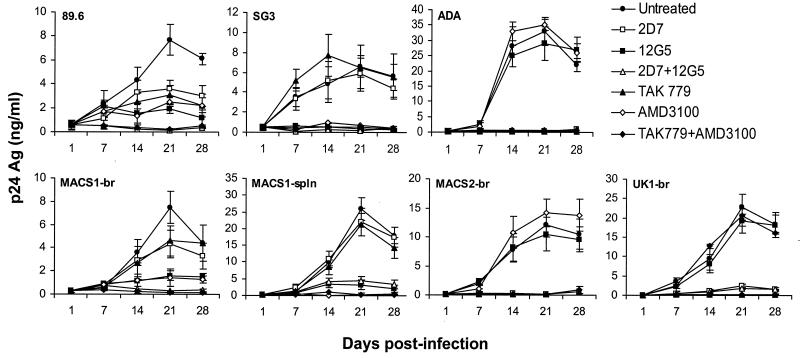
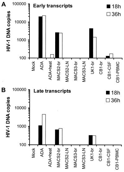
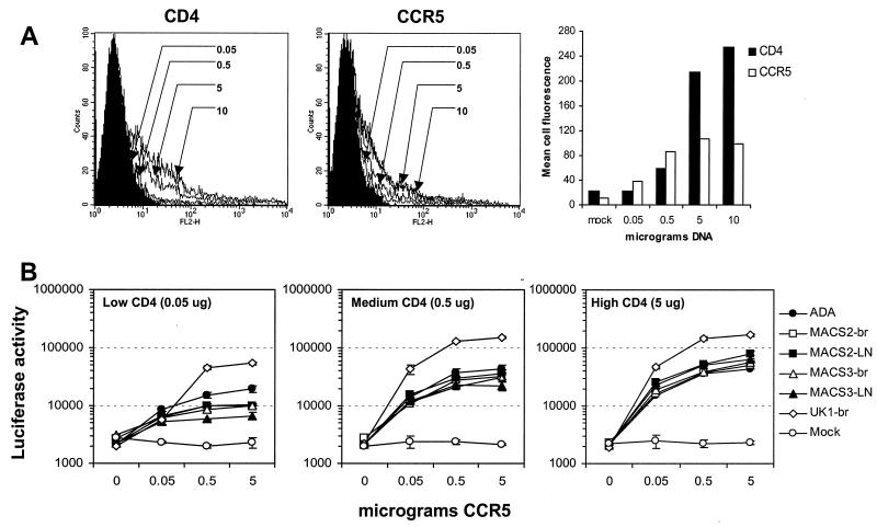
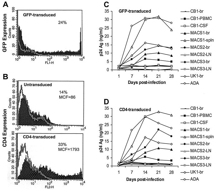
The viral determinants that underlie human immunodeficiency virus type 1 (HIV-1) neurotropism are unknown, due in part to limited studies on viruses isolated from brain. Previous studies suggest that brain-derived viruses are macrophage tropic (M-tropic) and principally use CCR5 for virus entry. To better understand HIV-1 neurotropism, we isolated primary viruses from autopsy brain, cerebral spinal fluid, blood, spleen, and lymph node samples from AIDS patients with dementia and HIV-1 encephalitis. Isolates were characterized to determine coreceptor usage and replication capacity in peripheral blood mononuclear cells (PBMC), monocyte-derived macrophages (MDM), and microglia. Env V1/V2 and V3 heteroduplex tracking assay and sequence analyses were performed to characterize distinct variants in viral quasispecies. Viruses isolated from brain, which consisted of variants that were distinct from those in lymphoid tissues, used CCR5 (R5), CXCR4 (X4), or both coreceptors (R5X4). Minor usage of CCR2b, CCR3, CCR8, and Apj was also observed. Primary brain and lymphoid isolates that replicated to high levels in MDM showed a similar capacity to replicate in microglia. Six of 11 R5 isolates that replicated efficiently in PBMC could not replicate in MDM or microglia due to a block in virus entry. CD4 overexpression in microglia transduced with retroviral vectors had no effect on the restricted replication of these virus strains. Furthermore, infection of transfected cells expressing different amounts of CD4 or CCR5 with M-tropic and non-M-tropic R5 isolates revealed a similar dependence on CD4 and CCR5 levels for entry, suggesting that the entry block was not due to low levels of either receptor. Studies using TAK-779 and AMD3100 showed that two highly M-tropic isolates entered microglia primarily via CXCR4. These results suggest that HIV-1 tropism for macrophages and microglia is restricted at the entry level by a mechanism independent of coreceptor specificity. These findings provide evidence that M-tropism rather than CCR5 usage predicts HIV-1 neurotropism.


Figures

FIG. 1
FIG. 1
V1/V2 and V3 HTA analysis. HIV-1 Env V1/V2 and V3 regions were amplified by RT-PCR of viral RNA and subjected to HTA analysis as described in Materials and Methods. (Left panels) V1/V2 and V3 heteroduplex patterns of HIV-1 isolated from brain and lymphoid tissue by coculture with CD8-depleted PBMC. (Right panels) V1/V2 and V3 heteroduplex patterns of HIV-1 isolated from the same tissues by coculture with CD8-depleted PBMC (lanes P) or MDM (lanes M). Virus isolates were from brain (br), lymph node (LN), or spleen (spln) tissues.
FIG. 2
FIG. 2
V1/V2 and V3 amino acid sequence analysis. The amino acid sequences were obtained from RT-PCR-amplified HIV-1 Env V1/V2 and V3 regions as described in Materials and Methods. V1/V2 and V3 alignments are compared to the clade B consensus sequence. Dots indicate residues identical to the clade B consensus, dashes indicate gaps, and X indicates uncertainty at the nucleotide level.
FIG. 3
FIG. 3
Coreceptor usage by primary HIV-1 isolates. Cf2-Luc cells were transfected with pcDNA3-CD4 alone or cotransfected with pcDNA3-CD4 and pcDNA3 expressing CCR2b, CCR3, CCR5, CCR8, CXCR4, CX3CR1, Gpr1, Gpr15, Strl33, or Apj and infected with equivalent amounts of each HIV-1 isolate as described in Materials and Methods. Viruses were isolated from brain (br), CSF, PBMC, lymph node (LN), spleen (spln), and spinal cord (SC) by coculture with CD8-depleted PBMC. Cell lysates were prepared at 48 h postinfection and assayed for luciferase activity. Data are expressed as means from duplicate infections. Error bars represent standard deviations. Similar results were obtained in two independent experiments.
FIG. 4
FIG. 4
Replication kinetics in PBMC. PBMC were infected with equivalent amounts of each virus, as described in Materials and Methods, and cultured for 18 days. Virus production in culture supernatants was measured by RT assays. Values shown are means from duplicate infections. Error bars represent standard deviations. Results are representative of two independent experiments using cells obtained from different donors.
FIG. 5
FIG. 5
Replication kinetics in MDM. MDM were infected with equivalent amounts of each virus, as described in Materials and Methods, and cultured for 27 days. Virus production in culture supernatants was measured by RT assays. Results are representative of two independent experiments using cells obtained from different donors.
FIG. 6
FIG. 6
Replication kinetics in microglia. Mixed brain cell cultures containing microglia were infected with equivalent amounts of each virus as described in Materials and Methods and cultured for 28 days. HIV-1 production in culture supernatants was measured by p24 antigen (Ag) ELISA (NEN). Data are represented as means from duplicate infections. Error bars represent standard deviations. Results are representative of three independent experiments using cells obtained from different donors.
FIG. 7
FIG. 7
Inhibition of HIV-1 infection in microglia by CCR5 and CXCR4 inhibitors. Mixed brain cell cultures containing microglia were treated with monoclonal antibody 2D7 (10 μg/ml), 12G5 (10 μg/ml), or both or treated with TAK779 (100 nM), AMD3100 (1.2 μM), or both for 1 h prior to infection. Untreated cells contained no antibody or inhibitor. Cells were infected with neurotropic primary isolates MACS1-br (R5X4), MACS1-spln (R5X4), MACS2-br (R5), and UK1-br (R5) or with control viruses 89.6 (R5X4), SG3 (X4), and ADA (R5) individually in the presence of each antibody or inhibitor as described in Materials and Methods and cultured for 28 days. HIV-1 production in culture supernatants was measured by p24 antigen (Ag) ELISA (NEN). Data are represented as means from duplicate infections. Error bars represent standard deviations. Results are representative of two independent experiments using cells obtained from different donors. The in vitro coreceptor usage phenotype for each virus determined with transfected Cf2-Luc cells (Fig. 3) is shown in parentheses.
FIG. 8
FIG. 8
Real-time PCR analysis of early (A) and late (B) viral transcripts in MDM. MDM were infected with equivalent amounts of DNase-treated virus stocks and analyzed by real-time PCR at 18 or 36 h after infection as described in Materials and Methods. Mock-infected cells were incubated with DNase-treated medium. ADA was heat inactivated by incubation at 56°C for 1 h. The results are expressed as copies of HIV-1 DNA per 10,000 copies of beta-globin. The data shown are representative of two independent experiments using cells obtained from different donors.
FIG. 9
FIG. 9
Effect of CD4 and CCR5 levels on infection by R5 viruses. (A) Cf2-Luc cells were transfected with 0.05, 0.5, 5, or 10 μg of CD4- or CCR5-expressing plasmid and analyzed for surface expression of CD4 or CCR5 by flow cytometry. The total amount of DNA in each transfection was adjusted to 10 μg with pCDNA3. (B) Cotransfected Cf2-Luc cells expressing low, medium, or high levels of CD4 (0.05, 0.5, and 5 μg of CD4-expressing plasmid, respectively) and low, medium, or high levels of CCR5 (0.05, 0.5, and 5 μg of CCR5-expressing plasmid, respectively) were infected with equivalent amounts of M-tropic (ADA, MACS2-br, and UK1-br) or non-M-tropic (MACS2-LN, MACS3-br, and MACS3-LN) R5 virus as described in Materials and Methods. Cell lysates were prepared at 48 h postinfection and assayed for luciferase activity. Data are expressed as means from duplicate infections. Error bars represent standard deviations. Similar results were obtained in two independent experiments.
FIG. 10
FIG. 10
Effect of high CD4 expression on virus replication in microglia. Microglia were untreated or transduced with retroviral vectors to express GFP (A) or CD4 (B). Microglia expressing high levels of CD4 (D) or GFP as a control (C) were infected with equivalent amounts of each HIV-1 isolate as described in Materials and Methods and cultured for 28 days. Virus production in culture supernatants was measured by p24 antigen (Ag) ELISA (NEN). Results are representative of those from two independent experiments with cells obtained from different donors. Percentages represent the proportion of cells expressing GFP or CD4. MCF, mean cell fluorescence.

References

    1. Albright A V, Shieh J T C, Itoh T, Lee B, Pleasure D, O'Connor M J, Doms R W, Gonzalez-Scarano F. Microglia express CCR5, CXCR4, and CCR3, but of these, CCR5 is the principal coreceptor for human immunodeficiency virus type 1 dementia isolates. J Virol. 1999;73:205–213. - PMC - PubMed
    1. Alkhatib G, Combadiere C, Broder C C, Feng Y, Kennedy P E, Murphy P M, Berger E A. CC CKR5: a RANTES, MIP-1α, MIP-1β receptor as a fusion cofactor for macrophage-tropic HIV-1. Science. 1996;272:1955–1958. - PubMed
    1. Arthos J, Rubbert A, Rabin R L, Cicala C, Machado E, Wildt K, Hanbach M, Steenbeke T D, Swofford R, Farber J M, Fauci A S. CCR5 signal transduction in macrophages by human immunodeficiency virus and simian immunodeficiency virus envelopes. J Virol. 2000;74:6418–6424. - PMC - PubMed
    1. Baba M, Nishimura O, Kanzaki N, Okamoto M, Sawada H, Iizawa Y, Shiraishi M, Aramaki Y, Okonogi K, Ogawa Y, Meguro K, Fujino M. A small molecule, nonpeptide CCR5 antagonist with highly potent and selective anti-HIV-1 activity. Proc Natl Acad Sci USA. 1999;96:5698–5703. - PMC - PubMed
    1. Balliet J W, Kolson D L, Eiger G, Kim F M, McGann K A, Srinivasan A, Collman R. Distinct effects in primary macrophages and lymphocytes of the human immunodeficiency virus type 1 accessory genes vpr, vpu, and nef: mutational analysis of a primary HIV-1 isolate. Virology. 1994;200:623–631. - PubMed

Publication types