. 2001 Nov;75(21):10326-33.
doi: 10.1128/JVI.75.21.10326-10333.2001.

cdc2 cyclin-dependent kinase binds and phosphorylates herpes simplex virus 1 U(L)42 DNA synthesis processivity factor

Affiliations

cdc2 cyclin-dependent kinase binds and phosphorylates herpes simplex virus 1 U(L)42 DNA synthesis processivity factor

S J Advani et al. J Virol. 2001 Nov.

Abstract

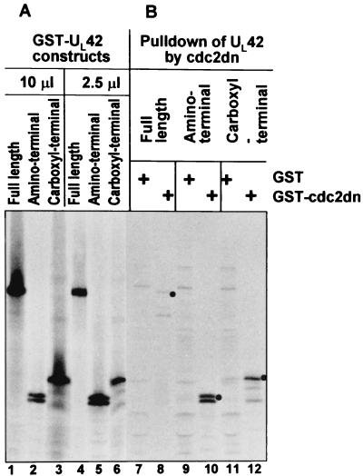
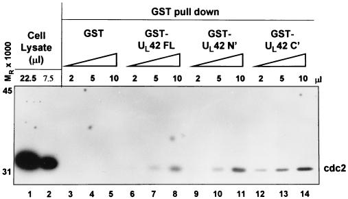
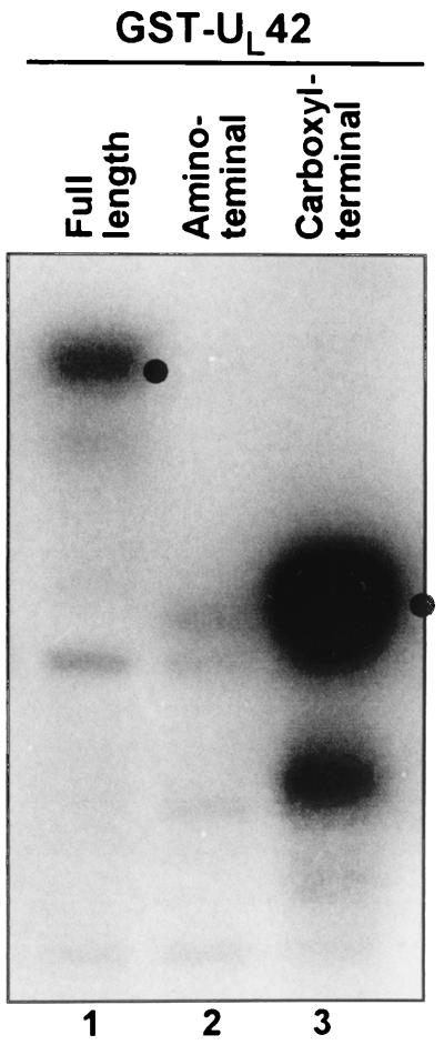
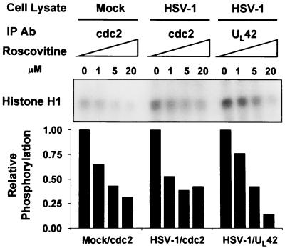
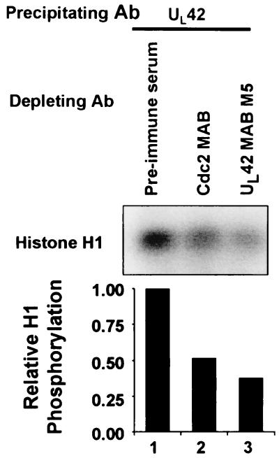
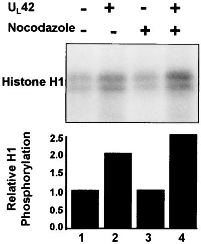
Earlier studies have shown that cdc2 kinase is activated during herpes simplex virus 1 infection and that its activity is enhanced late in infection even though the levels of cyclin A and B are decreased below levels of detection. Furthermore, activation of cdc2 requires the presence of infected cell protein no. 22 and the U(L)13 protein kinase, the same gene products required for optimal expression of a subset of late genes exemplified by U(S)11, U(L)38, and U(L)41. The possibility that the activation of cdc2 and expression of this subset may be connected emerged from the observation that dominant negative cdc2 specifically blocked the expression of U(S)11 protein in cells infected and expressing dominant negative cdc2. Here we report that in the course of searching for a putative cognate partner for cdc2 that may have replaced cyclins A and B, we noted that the DNA polymerase processivity factor encoded by the U(L)42 gene contains a degenerate cyclin box and has been reported to be structurally related to proliferating cell nuclear antigen, which also binds cdk2. Consistent with this finding, we report that (i) U(L)42 is able to physically interact with cdc2 at both the amino-terminal and carboxyl-terminal domains, (ii) the carboxyl-terminal domain of U(L)42 can be phosphorylated by cdc2, (iii) immunoprecipitates obtained with anti U(L)42 antibody contained a roscovitine-sensitive kinase activity, (iv) kinase activity associated with U(L)42 could be immunodepleted by antibody to cdc2, and (v) U(L)42 transfected into cells associates with a nocodazole-enhanced kinase. We conclude that U(L)42 can associate with cdc2 and that the kinase activity has the characteristic traits of cdc2 kinase.


Figures

FIG. 1
FIG. 1
Autoradiographic image of in vitro-transcribed/translated UL42 followed by GST-cdc2dn pull down. Full-length (FL) or amino (N′) or carboxyl (C′) halves of UL42 protein were in vitro transcribed and translated (lanes 1 to 6). The in vitro-translated UL42 proteins were then reacted with GST or GST–cdc2-dn for pull down assays (lanes 7 to 12), electrophoretically separated, and developed by autoradiography.
FIG. 2
FIG. 2
cdc2 immunoblot of HEp-2 cell lysates reacted with GST or GST fusion proteins expressing full-length (FL) or amino (N′) or carboxyl (C′) halves of UL42 protein. Immunoreactivity of cdc2 from whole-cell lysates is shown in lanes 1 and 2. Whole-cell lysates were then incubated with 2, 5, or 10 μl of GST (lanes 3 to 5), GST-UL42 FL (lanes 6 to 8), GST-UL42 N′ (lanes 9 to 11), or GST-UL42 C′ (lanes 12 to 14), and GST beads were collected, electrophoretically separated, and immunoblotted for cdc2.
FIG. 3
FIG. 3
Autoradiographic image of UL42 phosphorylation by cdc2 kinase. Full-length (lane 1), N-terminal (lane 2), C-terminal (lane 3) UL42 GST fusion proteins were incubated with purified cdc2 kinase in the presence of [γ-32P]ATP, separated by gel electrophoresis, and developed by autoradiography. Dots indicate the molecular weight of the respective GST fusion proteins.
FIG. 4
FIG. 4
Autoradiographic image of roscovitine-sensitive histone H1 phosphorylation by cdc2 or UL42 immunocomplexes. Mock- or HSV-1-infected HEp-2 cell lysates were subjected to immunoprecipitation (IP) by antibodies (Ab) to cdc2 or UL42. Immunocomplexes were preincubated with 0, 1, 5, or 20 μM roscovitine and subjected to in vitro kinase assays using histone H1 as the substrate. Phosphorylated histone 1 from the reactions was resolved by polyacrylamide gel electrophoresis, and the blots were developed by autoradiography. Histone phosphorylation was quantified by PhosphorImager analysis. Samples not treated roscovitine were assigned a value of 1.
FIG. 5
FIG. 5
Autoradiographic image of histone H1 kinase activity associated with UL42 immunocomplexes following immunodepletion. HSV-1-infected HEp2 cell lysates were immunodepleted with preimmune serum (lane 1), antibody (Ab) to cdc2 (lane 2), or antibody to UL42 (lane 3). The immunodepleted lysates were then immunprecipitated with antibody to UL42. Immunocomplexes were assayed for histone H1 kinase activity. Phosphorylated histone H1 was resolved by polyacrylamide gel electrophoresis, and the blots were developed by autoradiography. Histone phosphorylation was quantified by PhosphorImager analysis. The sample immunodepleted with preimmune serume was given a value of 1.
FIG. 6
FIG. 6
Autoradiographic image of histone H1 kinase activity from UL42 transfected cells. HEp-2 cells were transfected with pCDNA3.1 encoding UL42 (lanes 2 and 4). Cells were also treated with nocodazole following transfection (lanes 3 and 4). Cell lysates were then immunoprecipitated with antibody to UL42 and subjected to histone H1 kinase assays. Phosphorylated histone H1 was electrophoretically separated in a denaturing polyacrylamide gel and subjected to autoradiography. Histone phosphorylation was quantified by PhosphorImager analysis. Untransfected lysate without nocodazole treatment was assigned a value of 1.
FIG. 7
FIG. 7
Schematic diagram of cdc2 regulation and activity in the uninfected (top) and HSV-1-infected (bottom) cell. In uninfected cells, cdc2 is inactivated by wee-1 and myt-1 kinases and activated by cdc25C phosphatase, cyclin-dependent activating kinase (CAK), and association with cyclin B at the G2/M interphase. HSV-1 infection targets wee-1 and cdc25C through the actions of ICP22 and UL13, and UL42 associates with cdc2. The substrates for cdc2 in infected cells include ICP0, ICP4, and gI. The activation of cdc2 in the infected cell is associated with optimal accumulation of a subset of γ2 proteins exemplified by Us11 protein.

References

    1. Advani S J, Brandimarti R, Weichselbaum R R, Roizman B. The disappearance of cyclins A and B and the increase in activity of the G2/M-phase cellular kinase cdc2 in herpes simplex virus 1-infected cells require expression of the α22/US1.5 and UL13 viral genes. J Virol. 2000;74:8–15. - PMC - PubMed
    1. Advani S J, Weichselbaum R R, Roizman B. E2F protein are posttranslationally modified concomitantly with a reduction in nuclear binding activity in cells infected with herpes simplex virus 1. J Virol. 2000;74:7842–7850. - PMC - PubMed
    1. Advani S J, Weichselbaum R R, Roizman B. The role of cdc2 in the expression of herpes simplex virus genes. Proc Natl Acad Sci USA. 2000;97:10996–11001. - PMC - PubMed
    1. Advani S J, Hagglund R, Weichselbaum R R, Roizman B. Posttranslational processing of infected cell proteins 0 and 4 of herpes simplex virus 1 is sequential and reflects the subcellular compartment in which the proteins localize. J Virol. 2001;75:7904–7912. - PMC - PubMed
    1. Barrasa M I, Guo J T, Saputelli J, Mason W S, Seeger C. Does a cdc2 kinase-like recognition motif on the core protein of hepadnavirus regulate assembly and disintegration of capsids? J Virol. 2001;75:2024–2028. - PMC - PubMed

Publication types

LinkOut - more resources