. 2001 Nov;75(21):10472-8.
doi: 10.1128/JVI.75.21.10472-10478.2001.

The synthetic peptide P-197 inhibits human T-cell leukemia virus type 1 envelope-mediated syncytium formation by a mechanism that is independent of Hsc70

Affiliations

The synthetic peptide P-197 inhibits human T-cell leukemia virus type 1 envelope-mediated syncytium formation by a mechanism that is independent of Hsc70

D W Brighty et al. J Virol. 2001 Nov.

Abstract

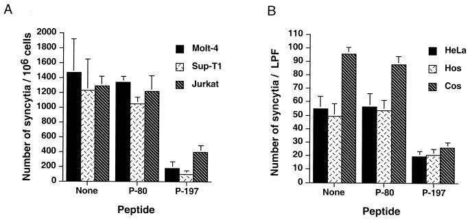
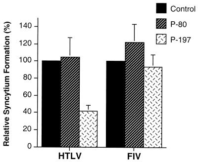
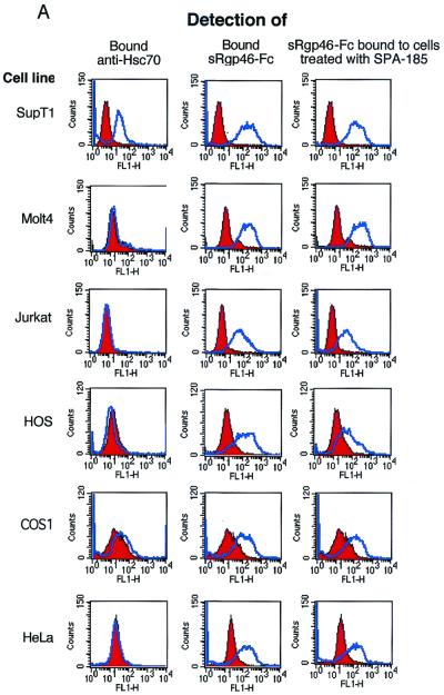
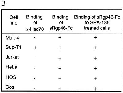
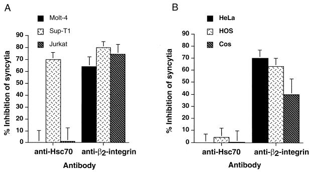
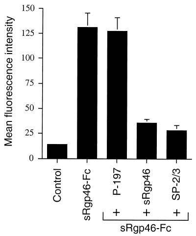
Entry of human T-cell leukemia virus type 1 (HTLV-1) into cells is mediated by the viral envelope glycoproteins gp46 and gp21. The gp46 surface glycoprotein binds to a poorly characterized cell surface receptor, thereby promoting the gp21-dependent fusion of the viral and cellular membranes. Interestingly, a synthetic peptide (P-197) simulating amino acids 197 to 216 of gp46 strongly inhibits envelope-dependent membrane fusion with Molt-4 target cells. It has been suggested that this peptide acts by competitively binding to Hsc70, a putative cellular receptor for HTLV-1. We now demonstrate that P-197 inhibits membrane fusion among diverse HTLV-1-permissive target cells. Importantly, most of these cells lack detectable levels of Hsc70, indicating that P-197 inhibits membrane fusion by a mechanism that is Hsc70 independent. We now suggest that competition for primary receptor binding is unlikely to account for the inhibitory activity of P-197. Understanding the mechanism by which P-197 functions may reveal concepts of general relevance to antiretroviral chemotherapy.


Figures

FIG. 1
FIG. 1
Peptide P-197 inhibits HTLV-1 envelope-mediated syncytium formation among diverse target cells. (A) Molt-4, Sup-T1, and Jurkat target cells were cocultured with HTLV-1-infected MT2 cells in the presence or absence of the indicated peptides (20 μg/ml). Control cocultures receiving no peptide were incubated in the presence of solvent only. The number of syncytia per 106 target cells was evaluated by high-power light microscopy. Data are means and standard deviations from six independent assays. (B) HeLa, HOS, or Cos target cells were cocultured with HTLV-1 envelope-expressing HeLa cells (transfected with the envelope expression vector pHTE-1) in the presence or absence of the indicated peptides. The number of syncytia per low-power field (LPF) was scored by light microscopy. Data are means and standard deviations from three fields from three independent assays.
FIG. 2
FIG. 2
The peptide P-197 does not block syncytium formation by an unrelated retrovirus. HeLa cells transfected with the HTLV-1 envelope expression construct HTE-1 (HTLV) or the feline immunodeficiency virus proviral clone pFIV-34TF10 (FIV) were cocultured with untransfected target HeLa cells in the presence of the indicated peptides. Syncytium formation is expressed as a percentage relative to the number of syncytia obtained with each viral envelope in the absence of peptide (Control). Data are means and standard deviations from three low-power fields from three independent assays.
FIG. 3
FIG. 3
Most cell lines permissive for HTLV-1 syncytium formation do not express Hsc70 on the cell surface. Binding of the anti-Hsc70 antibody SPA-815 (1 μg/ml) and the recombinant HTLV-1 SU derivative sRgp46-Fc (0.6 μg/ml) to target cells was monitored by flow cytometry. Binding of sRgp46-Fc (0.6 μg/ml) to cells in the presence of SPA-815 (4 μg/ml) is also shown. In each case the blue histograms represent basal fluorescence in the presence of irrelevant control sera and the red histograms represent the fluorescence profile in the presence of the detected test ligand. The data are summarized in panel B.
FIG. 3
FIG. 3
Most cell lines permissive for HTLV-1 syncytium formation do not express Hsc70 on the cell surface. Binding of the anti-Hsc70 antibody SPA-815 (1 μg/ml) and the recombinant HTLV-1 SU derivative sRgp46-Fc (0.6 μg/ml) to target cells was monitored by flow cytometry. Binding of sRgp46-Fc (0.6 μg/ml) to cells in the presence of SPA-815 (4 μg/ml) is also shown. In each case the blue histograms represent basal fluorescence in the presence of irrelevant control sera and the red histograms represent the fluorescence profile in the presence of the detected test ligand. The data are summarized in panel B.
FIG. 4
FIG. 4
Antibodies directed against Hsc70 do not block syncytium formation for most HTLV-1-permissive cells. (A) Molt-4, Sup-T1, and Jurkat cells were cocultured with HTLV-1-infected MT2 cells in the presence or absence of the indicated antibodies (2 μg/ml). The inhibition of syncytium formation relative to untreated control cocultures is indicated (means and standard deviations from four independent assays). (B) HeLa, HOS, and Cos target cells were cocultured with HTLV-1 envelope-expressing HeLa cells in the presence or absence of the indicated antibodies. The inhibition of syncytium formation relative to untreated control cocultures is indicated (means and standard deviations of triplicate determinations from three independent assays).
FIG. 5
FIG. 5
The peptide P-197 does not prevent binding of sRgp46-Fc to cells. Jurkat cells were treated with P-197 (40 μg/ml) and, for comparison, sRgp46 (0.6 μg/ml) or neutralizing anti-SU antiserum SP2-3 (1:1,000). sRgp46-Fc was added and incubated for 1 h with mixing. Bound sRgp46-Fc was detected using anti-Fc FITC-conjugated sera and flow cytometry. Basal fluorescence of control samples was determined from cells treated with irrelevant nonspecific IgG. Means and standard deviations from three independent assays are shown.

References

    1. Cann A J, Chen I S Y. Human T-cell leukemia virus type I and II. In: Fields B N, Knipe D M, Howley P M, editors. Fields virology. 3rd ed. Vol. 2. Philadelphia, Pa: Lippincott-Raven; 1996. pp. 1501–1527.
    1. Center R J, Kobe B, Wilson K A, Teh T, Howlett G J, Kemp B E, Poumbourios P. Crystallization of a trimeric human T-cell leukemia virus type 1 gp21 ectodomain fragment as a chimera with maltose-binding protein. Protein Sci. 1998;7:1612–1619. - PMC - PubMed
    1. Daenke S, McCracken S A, Booth S. Human T-cell leukaemia/lymphoma virus type 1 syncytium formation is regulated in a cell-specific manner by ICAM-1, ICAM-3 and VCAM-1 and can be inhibited by antibodies to integrin β2 or β7. J Gen Virol. 1999;80:1429–1436. - PubMed
    1. Delamarre L, Pique C, Pham D, Tursz T, Dokhelar M C. Identification of functional regions in the human T-cell leukemia virus type I SU glycoprotein. J Virol. 1994;68:3544–3549. - PMC - PubMed
    1. Delamarre L, Rosenberg A R, Pique C, Pham D, Dokhelar M C. A novel human T-leukemia virus type 1 cell-to-cell transmission assay permits definition of SU glycoprotein amino acids important for infectivity. J Virol. 1997;71:259–266. - PMC - PubMed

Publication types

MeSH terms

Substances

LinkOut - more resources