. 2001 Oct;19(10):946-51.
doi: 10.1038/nbt1001-946.

Quantitative profiling of differentiation-induced microsomal proteins using isotope-coded affinity tags and mass spectrometry

Affiliations

Quantitative profiling of differentiation-induced microsomal proteins using isotope-coded affinity tags and mass spectrometry

D K Han et al. Nat Biotechnol. 2001 Oct.

Abstract

An approach to the systematic identification and quantification of the proteins contained in the microsomal fraction of cells is described. It consists of three steps: (1) preparation of microsomal fractions from cells or tissues representing different states; (2) covalent tagging of the proteins with isotope-coded affinity tag (ICAT) reagents followed by proteolysis of the combined labeled protein samples; and (3) isolation, identification, and quantification of the tagged peptides by multidimensional chromatography, automated tandem mass spectrometry, and computational analysis of the obtained data. The method was used to identify and determine the ratios of abundance of each of 491 proteins contained in the microsomal fractions of naïve and in vitro- differentiated human myeloid leukemia (HL-60) cells. The method and the new software tools to support it are well suited to the large-scale, quantitative analysis of membrane proteins and other classes of proteins that have been refractory to standard proteomics technology.


Figures
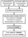
Figure 1
Figure 1
Schematic representation of the quantitative proteomics procedure. Proteins in the microsomal fraction of naïve or PMA-treated HL-60 cells were labeled with ICAT reagents, combined, and analyzed as described in the text.
Figure 2
Figure 2
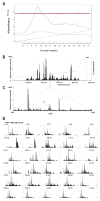
Multidimensional liquid chromatography tandem mass spectrometric analysis of a complex peptide mixture. (A) Distribution of peptides contained in tryptic-digested HL-60 microsomal fraction on a strong cation-exchange chromatography column. Peptides were detected by absorbance at 214 nm (blue line) and 280 nm (green line). Solvent gradient (red line), and pressure (pink line) are also indicated. Collected fraction numbers are shown on the x-axis. (B) Analysis of the biotinylated, cysteine-containing peptides contained in cation-exchange fraction 18 by μLC-ESI-MS/MS. Ion chromatogram displaying the base peak (most intense ion signal in each MS scan) as a function of retention time. Dotted line indicates the percentage acetonitrile solvent gradient used to develop the reverse-phase capillary column. (C) MS spectrum of peptides detected in the 30 s time window indicated in (B). Signals indicated with asterisk (*) were ICAT-labeled peptides that were subjected to automated CID. The data obtained from the peptides numbered as 1 and 2 are shown in Figure 3. (D) Base peak ion chromatogram of all of the cation-exchange fractions collected, indicating ICAT peptide distribution. Red line indicates solvent gradient.
Figure 3
Figure 3
Post-MS processing of data from selected peptides. Two peptides numbered as 1 (A) and 2 (B) in Figure 2C were subjected to alternating MS and MS/MS scans. For each peptide we show the CID spectrum, the identity of the parent protein determined by sequence database searching using SEQUEST (C* designates cysteine labeled with the heavy form of ICAT reagent, while C# designates cysteine labeled with the light form of ICAT reagent), the data indicating the relative abundance, and the calculated d0:d8 ratio obtained using XPRESS software. (C) Flowchart indicating the steps applied to process the initial CID spectra to identify and quantify microsomal proteins.
Figure 4
Figure 4
Consistency of redundant protein quantification. A total of 13 peptides from the CD45 transmembrane protein tyrosine phosphatase CD45 were identified and quantified. (A) CD45 protein sequence with the identified, ICAT reagent-labeled peptides indicated in boxes with yellow background. (B) Redundancy and accuracy of quantitation: the amino acid sequence of the identified peptides, the cation-exchange fraction(s) in which each peptide was detected, the number of times each peptide was identified and quantified, and the abundance ratio and its standard deviations are indicated. Results are shown from three independent experiments: one large scale (first experiment) and two smaller scale (second and third experiments).
Figure 5
Figure 5
Categories of proteins identified in this study. The 491 proteins identified and quantified in this study were classified by broad functional criteria. The numbers in parentheses indicate the percentage fraction of identified proteins represented by each category. Some proteins are represented in more than one category (e.g., CD45 was counted as both a transmembrane protein and a protein phosphatase). Protein sequences from hypothetical proteins (81% containing possible transmembrane α-helices) and functionally uncharacterized proteins (78% containing possible transmembrane α-helices) were analyzed for the possible presence of transmembrane helices using the Tmpred tool available on EXPASY website (http://www.ch.embnet.org/software/TMPRED_form.html).

References

    1. Oseroff AR, Robbins PW, Burger MM. Cell surface membrane: biochemical aspects and biophysical probes. Annual Rev Biochem. 1973;42:647–682. - PubMed
    1. Liu E, et al. The HER2 (c-erbB-2) oncogene is frequently amplified in in situ carcinomas of the breast. Oncogene. 1992;7:1027–1032. - PubMed
    1. Shak S. Overview of the trastuzumab (Herceptin) anti-HER2 monoclonal antibody clinical program in HER2-overexpressing metastatic breast cancer. Herceptin Multinational Investigator Study Group. Semin Oncol. 1999;26:71–77. - PubMed
    1. Eccles SA. Monoclonal antibodies targeting cancer: “magic bullets” or just the trigger? Breast Cancer Res. 2001;3:86–90. - PMC - PubMed
    1. Drews J. Research and development. Basic science and pharmaceutical innovation. Nat Biotechnol. 1999;17:406–408. - PubMed

Publication types