. 2001 Oct;159(4):1313-21.
doi: 10.1016/S0002-9440(10)62518-7.

Renal fibrosis: collagen composition and assembly regulates epithelial-mesenchymal transdifferentiation

Affiliations

Renal fibrosis: collagen composition and assembly regulates epithelial-mesenchymal transdifferentiation

M Zeisberg et al. Am J Pathol. 2001 Oct.

Abstract

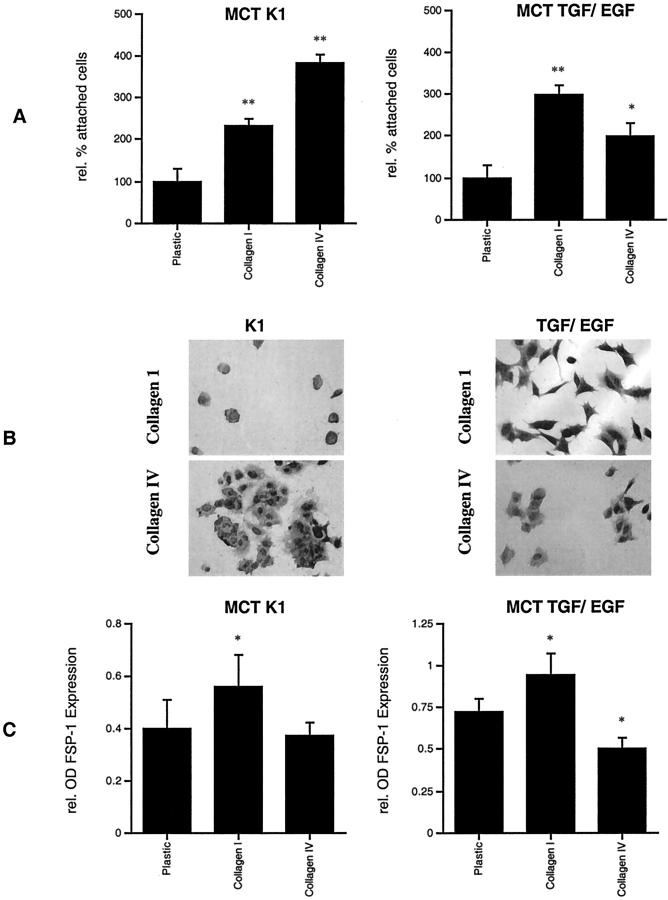
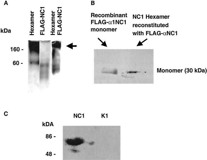
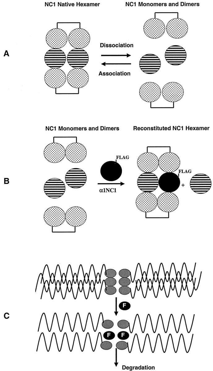
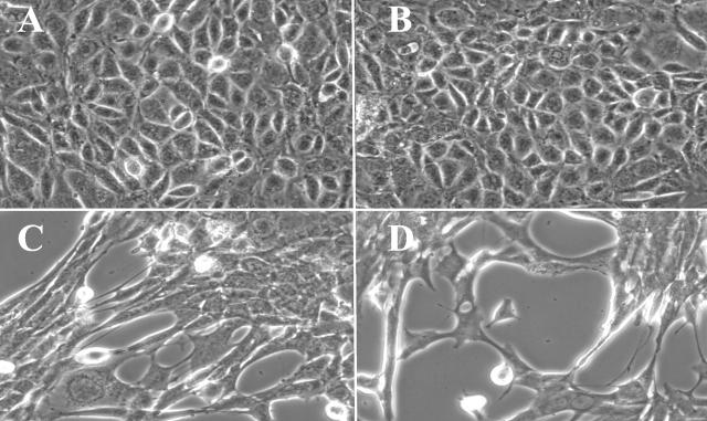
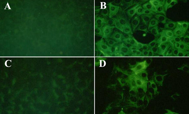
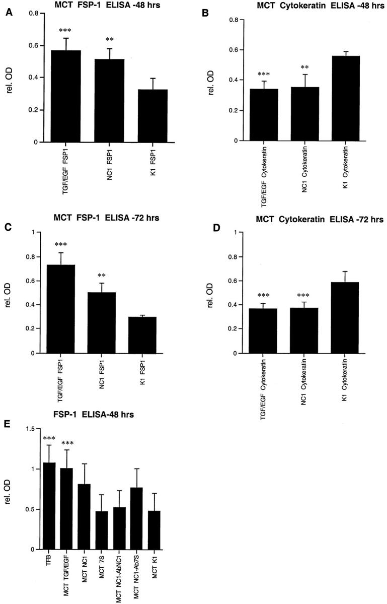
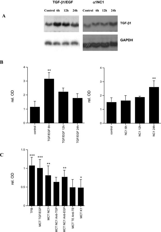
Type IV collagen is a major component of basement membranes and it provides structural and functional support to various cell types. Type IV collagen exists in a highly complex suprastructure form and recent studies implicate that protomer (the trimeric building unit of type IV collagen) assembly is mediated by the NC1 domain present in the C-terminus of each collagen alpha-chain polypeptide. Here we show that type IV collagen contributes to the maintenance of the epithelial phenotype of proximal tubular epithelial cells, whereas type I collagen promotes epithelial-to-mesenchymal transdifferentiation (EMT). In addition, the recombinant human alpha1NC1 domain inhibits assembly of type IV collagen NC1 hexamers and potentially disrupts the deposition of type IV collagen, facilitating EMT in vitro. Inhibition of type IV collagen assembly by the alpha1NC1 domain up-regulates the production of transforming growth factor-beta1 in proximal tubular epithelial cells, an inducer of EMT. These results strongly suggest that basement membrane architecture is pivotal for the maintenance of epithelial phenotype and that changes in basement membrane architecture potentially lead to up-regulation of transforming growth factor-beta1, which contributes to EMT during renal fibrosis.


Figures

Figure 1.
Figure 1.
Tubular epithelial cell interactions with collagen types I and IV. MCT cells adhere preferably to type IV collagen (383 ±16.6% compared to uncoated plastic control) than to type I collagen (232.2 ± 20.5%) in cell adhesion assay (A, left). After induction of EMT with TGF-β1 and EGF, MCT cells with a fibroblast-like morphology attached increasingly to collagen type I (396.7 ± 24.3% compared to uncoated plastic control), whereas adhesion to type IV collagen was less abundant (299.5 ± 20.6%) (A, right). MCT cells grown in K1 medium adhere strongly to type IV collagen and seem to display a round-shaped, epithelial cell-like, morphology (B, left). MCT cells that were pretreated with TGF-β1 and EGF gain in capacity to attach to type I collagen and appear to have a more spindle-shaped morphology (B, right). Cultivation on type I collagen increased FSP-1 expression in MCT cells that were grown in K1 medium (140 ± 9.3% compared to uncoated plastic control), whereas cultivation on type IV collagen had no effect on FSP-1 expression in untreated cells (C, left) as was measured by ELISA of cell lysates. When EMT was induced in MCT cells with TGF-β1 and EGF, cultivation on type I collagen further increased levels of FSP-1 expression (130.6 ± 7.3% compared to uncoated plastic control), whereas coating with type IV collagen decreased levels of FSP-1 expression (58.1 ± 10.4%) and thus stabilized the epithelial phenotype (C, right). *, P < 0.05; **, P < 0.001. Original magnifications, ×400.
Figure 2.
Figure 2.
Recombinant α1NC1 domain is incorporated into hexamers of native NC1 protein. To demonstrate incorporation of recombinant FLAG-α1NC1 domain into hexamers, native hexamers of type IV collagen from kidney cortex were subjected to changing pH from 7.5 to 3.0 and subsequently allowed to reassemble in the presence of FLAG-α1NC1 monomers. Native hexamers and FLAG-α1NC1 display typical bands (A). When hexamers were subjected to changing of pH and allowed to reassemble in the presence of the FLAG-α1NC1 domain and were run on a denaturing gel FLAG-α1NC1 incorporated into hexamers. Bands corresponding to the full-size hexamer were cut out (A, arrow). Protein was extracted and subjected to dissociation by SDS-PAGE. Western Blot analysis with anti-FLAG antibody revealed positive staining of eluted protein (B) and thus proves incorporation of FLAG-α1NC1 domain into hexamers. When FLAG-α1NC1 domain was added to MCT cells, Western blot analysis of cell culture supernatant with antibodies to type IV collagen revealed an increased pattern that is typical of type IV collagen degradation (C, left lane) in contrast to untreated cells (C, right lane). Thus it demonstrates an increased disassembly of type IV collagen in cell culture.
Figure 3.
Figure 3.
Schematic illustration of incorporation of recombinant FLAG-α1NC1 domain into type IV collagen hexamers and its potential capacity to degrade type IV collagen. When type IV collagen hexamers are subjected to low pH treatment they dissociate to monomers and dimers (A). When NC1 monomers and dimers are reassembled into hexamers in the presence of FLAG-tagged recombinant α1NC1 domain, it gets incorporated into the hexameric structure (B). Because of the lack of collagenous domain and 7S domain, incorporation of FLAG-α1NC1 domain potentially leads to increased degradation of type IV collagen (C).
Figure 4.
Figure 4.
MCT cells acquire a fibroblast-like morphology after incubation with type IV collagen α1NC1 domain, similar to changes that were observed after incubation with TGF-β1 and EGF. Tubular epithelial MCT cells display their typical cobblestone-like morphology when cultured in K1 medium (A). When medium was supplemented with either soluble type IV collagen α1NC1 (C) domain or with 3 ng/ml TGF-β1 and 10 ng/ml EGF (D) cells acquired a typical spindle-shaped morphology, which is typically observed in the induction of EMT. Incubation with type IV collagen 7S domain did not alter the cellular phenotype (B). Original magnifications, ×400.
Figure 5.
Figure 5.
Incubation with type IV collagen α1NC1 domain results in increased expression of mesenchymal markers FSP-1 and vimentin by MCT cells. MCT cells that were cultured in K1 medium displayed little staining for the mesenchymal markers vimentin (A) and FSP-1 (C) using indirect immunofluorescent staining. Treatment with type IV collagen α1NC1 domain resulted in a robust up-regulation of vimentin (B) and FSP-1 (D) expression. Original magnifications, ×400.
Figure 6.
Figure 6.
Solid phase direct ELISA for FSP-1 and cytokeratin. Treatment of MCT cells with type IV collagen α1NC1 domain resulted in an increase in FSP-1 expression (A and C) and a decrease in cytokeratin expression as compared to untreated K1 control (B and D). Effects were similar to those that were obtained by stimulation with TGF-β1 and EGF, which is currently the most established inductor of EMT in vitro. After 48 hours the α1NC1 domain induced an increase in FSP-1 expression (162.7 ± 14.1% compared to K1 control) (A) and decreased cytokeratin expression (76.1 ± 5.3% compared to K1 control) (B), whereas effects of TGF-β1 and EGF were 156.7 ± 23.6% and 65.0 ± 4.9%, respectively. Similar results were obtained after 72 hours (C and D). Type IV collagen α1NC1 domain induced FSP-1 expression (181.6 ± 23.6% compared to control) (C) and a decrease in cytokeratin expression by (61.4 ± 9.6% of K1 control) (D). Treatment with TGF-β1 and EGF for 72 hours resulted in an induction of FSP-1 expression (184.7 ± 28.1%) (C) and depression of cytokeratin expression (60.7 ± 10.1%) (D). In another series of experiments FSP-1 expression was determined by ELISA to test the specificity of α1NC1 domain-induced effects (E). ELISAs for FSP-1 display a strong expression in tubulointerstitial fibroblasts and low expression levels in untreated MCT cells (MCT-K1) that were used as a control. Increased expression of FSP-1 compared to untreated MCT control was detectable after treatment with TGF-β1 and EGF (259.9 ± 40.2% compared to MCT K1 control) as well as with α1NC1 domain (199.2 ± 33.1%). Incubation with type IV collagen 7S domain had no significant effect on FSP-1 expression levels (105.9 ± 1.4%). Co-incubation of α1NC1 domain with α1NC1 neutralizing antibodies (MCT NC1-abNC1) diminished the increase in FSP-1 expression (112.7 ± 7.2%), whereas 7S neutralizing antibodies (MCT NC1-ab7S) had no effect (184.1 ± 23.7%). *, P < 0.05; **, P < 0.001; ***, P < 0.0001.
Figure 7.
Figure 7.
TGF-β1 mRNA expression is up-regulated in EMT. Induction of EMT by TGF-β1/EGF or with type IV collagen α1NC1 domain results in up-regulation of TGF-β1 mRNA expression (A). Induction with TGF-β1/EGF resulted in a peak after 6 hours, whereas after incubation with type IV collagen α1NC1 domains resulted in an up-regulation after 24 hours (B) summarizes the densitometric analysis of experiments with TGF-β1/EGF (left) and with α1NC1 domain (right). Treatment of MCT cells with TGF-β1 and EGF results in an increase in TGF-β1 mRNA after 6 hours by (291 ± 48% compared to K1 control), and remains elevated after 12 hours (204 ± 34%) and 24 hours (162 ± 18%). Treatment with α1NC1 domain leads to a significant increase in TGF-β1 mRNA after 24 hours (175.2 ± 24%), however results after 6 hours (110.7 ± 8%) and 12 hours (125.7 ± 9%) were not significant. In solid-phase direct ELISA for FSP-1 (C) co-incubation of α1NC1 domain with neutralizing antibodies to TGF-β1 (MCT NC1 anti-TGF) reduced the increase in FSP-1 expression significantly (158.1 ± 23% compared to K1 control), whereas the addition of neutralizing EGF antibodies (MCT NC1 anti-EGF) had no significant effect (193.5 ± 29%). Addition of neutralizing antibodies to TGF and EGF (MCT TE anti-TE) abolished growth factor-induced EMT (111.8 ± 14%). These results suggest a role for TGF-β1 autocrine stimulation for mediation of EMT. *, P < 0.05); **, P < 0.001; ***, P < 0.0001.

Comment in

References

    1. Yurchenco PD, O’Rear JJ: Basal lamina assembly. Curr Opin Cell Biol 1994, 6:674-681 - PubMed
    1. Hudson BG, Reeders ST, Tryggvason K: Type IV collagen: structure, gene organization, and role in human diseases. Molecular basis of Goodpasture and Alport syndromes and diffuse leiomyomatosis. J Biol Chem 1993, 268:26033-26036 - PubMed
    1. Timpl R, Oberbaumer I, von der Mark H, Bode W, Wick G, Weber S, Engel J: Structure and biology of the globular domain of basement membrane type IV collagen. Ann NY Acad Sci 1985, 460:58-72 - PubMed
    1. Yurchenco PD: Assembly of basement membranes. Ann NY Acad Sci 1990, 580:195-213 - PubMed
    1. Kalluri R, Cosgrove D: Assembly of type IV collagen. Insights from alpha3(IV) collagen-deficient mice. J Biol Chem 2000, 275:12719-12724 - PubMed

Publication types