. 2001 Oct;159(4):1335-43.
doi: 10.1016/s0002-9440(10)62520-5.

Aortic valve endothelial cells undergo transforming growth factor-beta-mediated and non-transforming growth factor-beta-mediated transdifferentiation in vitro

Affiliations

Aortic valve endothelial cells undergo transforming growth factor-beta-mediated and non-transforming growth factor-beta-mediated transdifferentiation in vitro

G Paranya et al. Am J Pathol. 2001 Oct.

Abstract

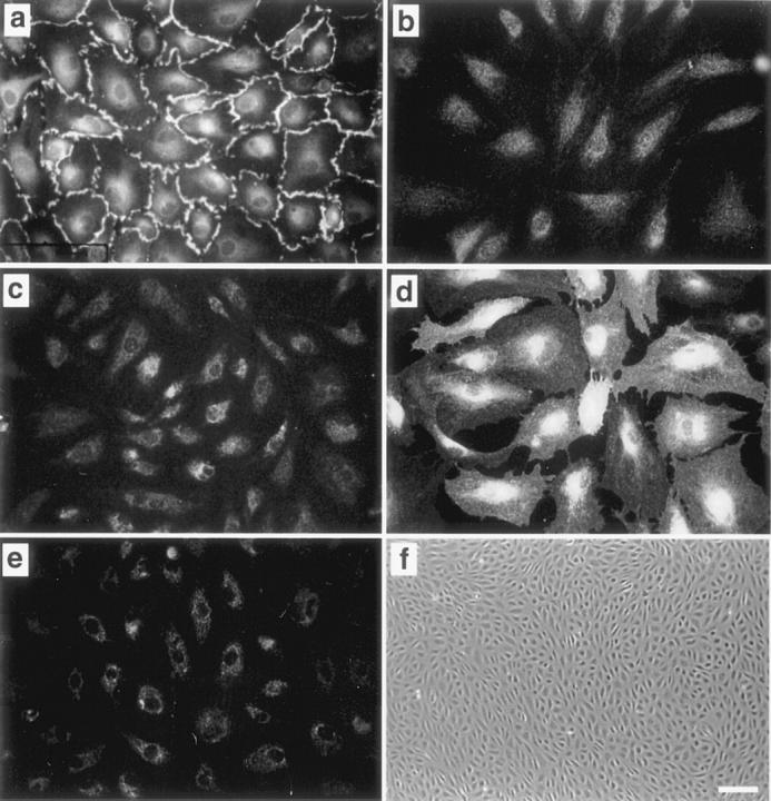
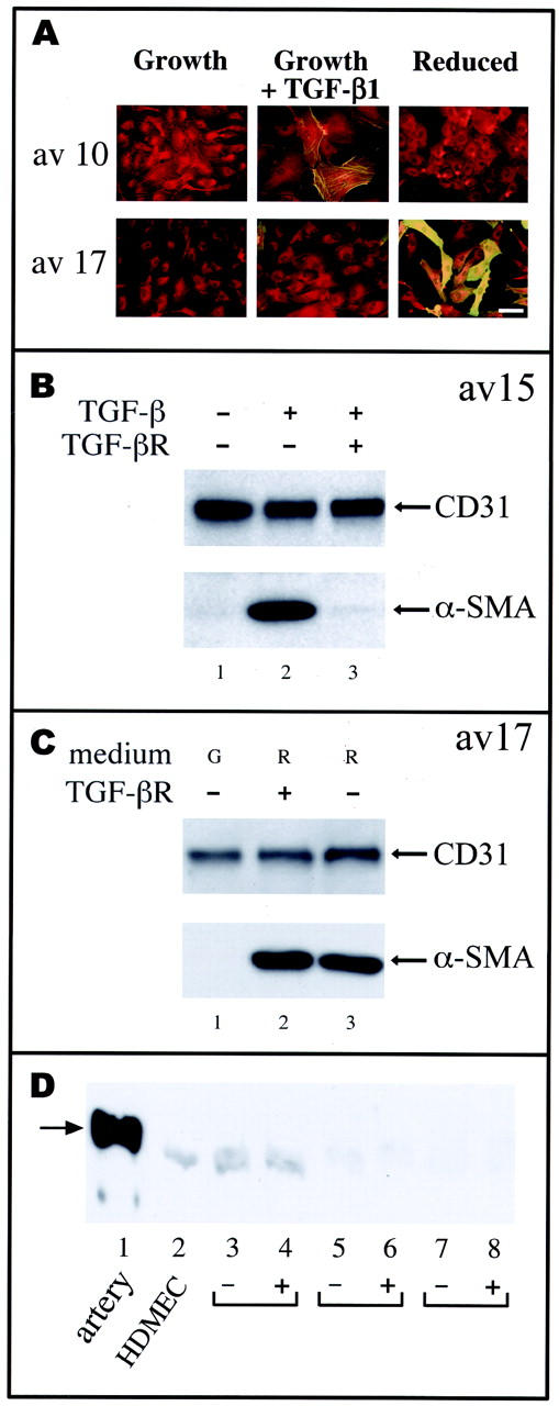
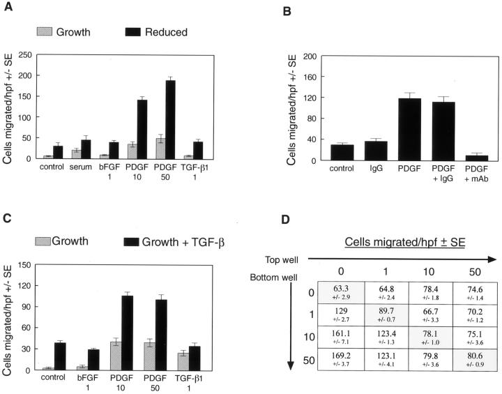
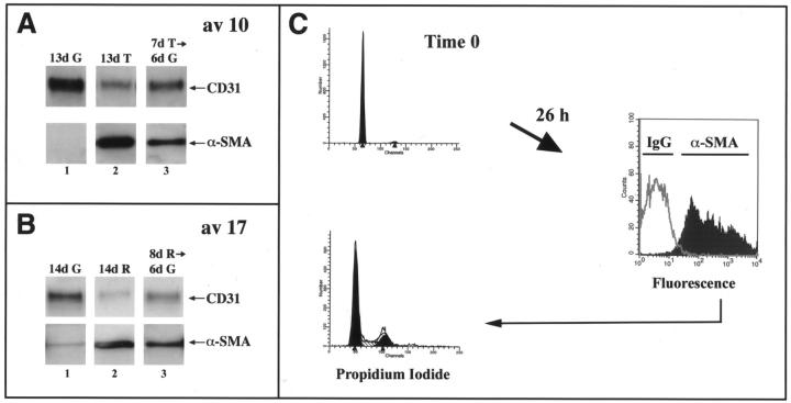
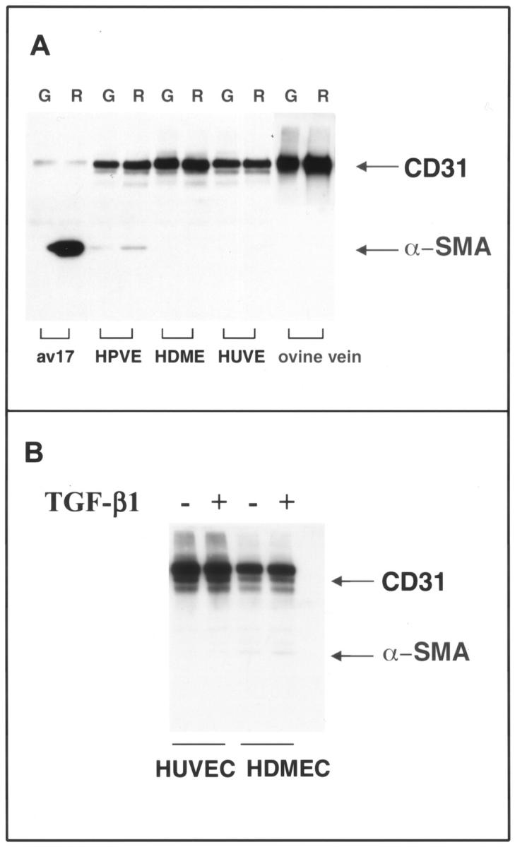
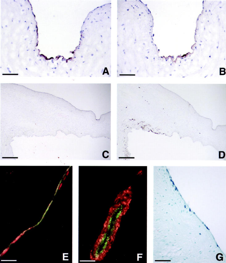
Cardiac valves arise from endocardial cushions, specialized regions of the developing heart that are formed by an endothelial-to-mesenchymal cell transdifferentiation. Whether and to what extent this transdifferentiation is retained in mature heart valves is unknown. Herein we show that endothelial cells from mature valves can transdifferentiate to a mesenchymal phenotype. Using induction of alpha-smooth muscle actin (alpha-SMA), an established marker for this process, two distinct pathways of transdifferentiation were identified in clonally derived endothelial cell populations isolated from ovine aortic valve leaflets. alpha-SMA expression was induced by culturing clonal endothelial cells in medium containing either transforming growth factor-beta or low levels of serum and no basic fibroblast growth factor. Cells induced to express alpha-SMA exhibited markedly increased migration in response to platelet-derived growth factor-BB, consistent with a mesenchymal phenotype. A population of the differentiated cells co-expressed CD31, an endothelial marker, along with alpha-SMA, as seen by double-label immunofluorescence. Similarly, this co-expression of endothelial markers and alpha-SMA was detected in a subpopulation of cells in frozen sections of aortic valves, suggesting the transdifferentiation may occur in vivo. Hence, the clonal populations of valvular endothelial cells described here provide a powerful in vitro model for dissecting molecular events that regulate valvular endothelium.


Figures

Figure 1.
Figure 1.
Clonal cells from ovine aortic valve express endothelial-specific markers. Av 10 cells were incubated with anti-human CD31 (a), control serum (b), anti-bovine E-selectin (c and d), or mouse anti-α-SMA (e). d: Av 10 cells were treated with 1 μg/ml lipopolysaccharide for 5 hours to induce E-selectin expression. f: Phase contrast micrograph of clonal aortic valve ECs. Original magnifications: ×400 (a–e), ×40 (f). Scale bar in f, 100 μm.
Figure 2.
Figure 2.
TGF-β-dependent and TGF-β-independent pathways for induction of α-SMA. A: Clones av 10 and av 17 were grown for 6 days in growth medium (growth, G), in growth medium with 1 ng/ml TGF-β1 (growth plus TGF-β1, T), or in medium with 0.5% serum and without bFGF (reduced, R). Cells were double-labeled with anti-CD31 and a Texas Red-conjugated secondary antibody and with anti-α-SMA and fluorescein-conjugated anti-mouse IgG for simultaneous detection of these two antigens. Original magnification, ×400. Scale bar, 10 μm. B: Clone av 15 cells cultured in growth medium (lane 1), in growth medium with 1 ng/ml TGF-β1 (lane 2), or in growth medium with TGF-β1 plus 36 μg/ml recombinant soluble TGF-β type II receptor antagonist (lane 3) for 6 days. C: Clone av 17 cells were cultured in growth medium (G) (lane 1), in reduced medium (R) with 36 μg/ml soluble TGF-β receptor (lane 2), and in reduced medium (R) (lane 3). Cell lysates were analyzed by Western blot for expression of CD31 and α-SMA. D: Lysates from ovine carotid artery (lane 1), cultured HDMECs (lane 2), aortic valve clones av 14 (lanes 3 and 4), av 17 (lanes 5 and 6), and av 10 (lanes 7 and 8) were probed by Western blot using anti-smooth muscle myosin heavy chain SM-1 monoclonal antibody. Av clones were in growth medium (−) or in reduced medium (+) for 7 days. The arrow denotes the 204-kd smooth muscle myosin heavy chain detected in ovine carotid artery. The faint band detected in HDMECs and aortic valve clones was designated nonspecific because of its presence in cultured ECs.
Figure 3.
Figure 3.
Transdifferentiated cells exhibit increased migration in response to PDGF-BB. A: Av 17 cells cultured in growth medium (hatched bars) or in reduced medium (solid bars) for 6 days were tested for the ability to migrate toward EBM with 0.1% bovine serum albumin (control), EBM with 5% FBS (serum), 1 ng/ml bFGF, 10 ng/ml PDGF-BB, 50 ng/ml PDGF-BB, or 1 ng/ml TGF-β1. B: A neutralizing anti-PDGF-BB monoclonal antibody (mAb) and an isotype-matched IgG were tested for the ability to block the migration of av 17 cells, which were grown in reduced medium for 6 days, toward 10 ng/ml PDGF-BB. C: Av 15 cells cultured in growth medium (hatched bars) or in growth medium with 1 ng/ml TGF-β1 (solid bars) for 6 days were tested as in A. D: Checkerboard analysis was performed on av 17 cells grown in reduced medium for 6 days. PDGF-BB at 0, 1, 10, or 50 ng/ml was added to top and bottom wells as indicated, and cells allowed to migrate for 4 hours. The shaded boxes on the diagonal highlight the finding that PDGF-BB does not elicit random cell migration toward increasing concentrations of PDGF-BB in the upper and lower chambers.
Figure 4.
Figure 4.
Characterization of endothelial transdifferentiation. A: Av 10 cells were cultured in growth medium (G) (lane 1) or in growth medium with 1 ng/ml of TGF-β1 (T) for 7 days (lanes 2 and 3). After 7 days, cells were trypsinized and replated on new gelatin-coated dishes in growth medium (lanes 1 and 3) or in growth medium with TGF-β1 (lane 2) for 6 additional days. B: A similar time series was used with av 17 cells and reduced medium (R) instead of TGF-β. Cell lysates were analyzed by Western blot using anti-CD31 and anti-α-SMA. C: Av 17 cells were grown in reduced medium for 6 days. A portion of the cells was stained with propidium iodide to determine the cell-cycle distribution (top left). The rest of the cells were replated in growth medium containing 10% FBS and 2 ng/ml of bFGF at 30,000 cells/cm for 26 hours. The cells were then harvested, permeabilized with 0.1% Triton-X-100 for immunostaining of intracellular α-SMA by flow cytometry (right), and simultaneously stained with propidium iodide (bottom left). The gray line at the right represents cells stained with an isotype-matched IgG2a control.
Figure 5.
Figure 5.
Induction of α-SMA is restricted to valvular ECs. A: Human pulmonary valve ECs (HPVEs), HDMECs, HUVECs, and ovine peripheral vein ECs (ovine vein) were grown for 6 days in growth medium (G) or in reduced medium (R). Av 17 cells served as a positive control. B: HDMECs and HUVECs were cultured in the absence or presence of 1 ng/ml TGF-β1 for 6 days. For both A and B, cell lysates were analyzed by Western blot using anti-CD31 and anti-α-SMA.
Figure 6.
Figure 6.
Transdifferentiation in mature valve leaflets. Immunoperoxidase staining of adjacent frozen sections of ovine aortic valve with anti-CD31 (A and C) and anti-α-SMA (B and D) is shown at ×400 original magnification (A and B) to focus on regions of co-localization of the two antigens and ×40 original magnification (C and D). The indented region seen on the upper surface of the leaflet on the right of C and D corresponds to the region shown in A and B. Scale bars: 12.5 μm (A and B) and 125 μm (C and D). E and F: Double-label immunofluorescence of ovine aortic valve leaflets with anti-vWF (green) and anti-α-SMA (red). F: A double-labeled vessel located toward the base of the leaflet as an internal control. G: A paraffin section of human aortic valve leaflet stained with anti-α-SMA at ×100 original magnification. Scale bar, 50 μm.

References

    1. Eisenberg LM, Markwald RR: Molecular regulation of atrioventricular valvuloseptal morphogenesis. Circ Res 1995, 77:1-6 - PubMed
    1. Schoen FJ, Levy RJ: Tissue heart valves: current challenges and future research perspectives. J Biomed Mat Res 1999, 47:439-465 - PubMed
    1. Mjaatvedt CH, Yamamura H, Wessels A, Ramsdell A, Turner D, Markwald RR: Mechanisms of segmentation, septation, and remodeling of the tubular heart: endocardial cushion fate and cardiac looping. Harvery RP Rosenthal N eds. Heart Development. 1999, :pp 159-177 Academic Press, San Diego
    1. Ramsdell AF, Markwald RR: Induction of endocardial cushion tissue in the avian heart is regulated in part by TGF-beta3-mediated autocrine signalling. Dev Biol 1997, 188:64-74 - PubMed
    1. Boyer AS, Ayerinskas II, Vincent EB, McKinney LA, Weeks DL, Runyan RB: TGF beta 2 and TGF beta 3 have separate and sequential activities during epithelial-mesenchymal cell transformation in the embryonic heart. Dev Biol 1999, 208:530-545 - PubMed

Publication types

Substances

LinkOut - more resources