. 2001 Oct;159(4):1397-404.
doi: 10.1016/S0002-9440(10)62526-6.

Suppression by apoptotic cells defines tumor necrosis factor-mediated induction of glomerular mesangial cell apoptosis by activated macrophages

Affiliations

Suppression by apoptotic cells defines tumor necrosis factor-mediated induction of glomerular mesangial cell apoptosis by activated macrophages

J S Duffield et al. Am J Pathol. 2001 Oct.

Abstract

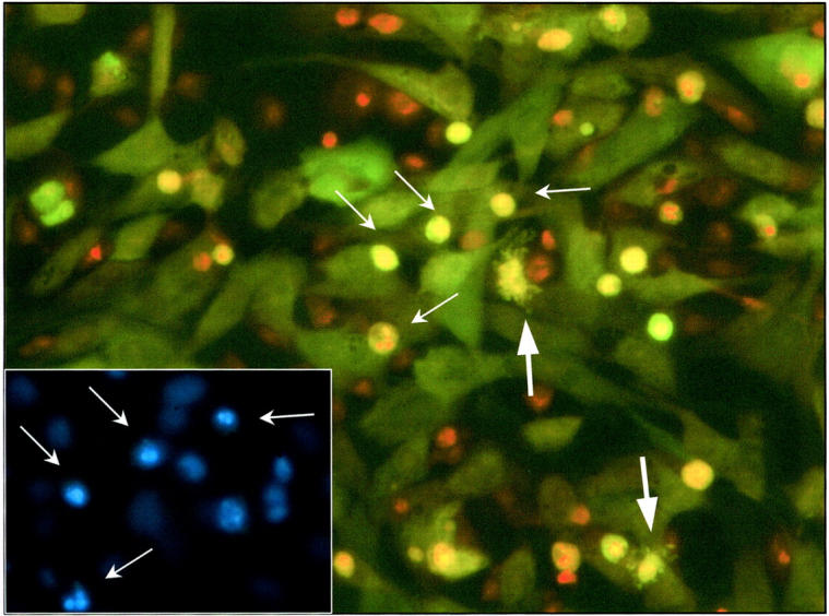
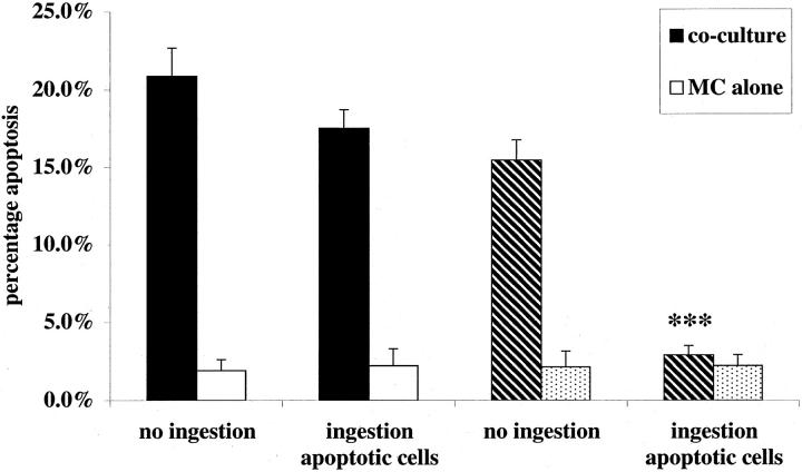
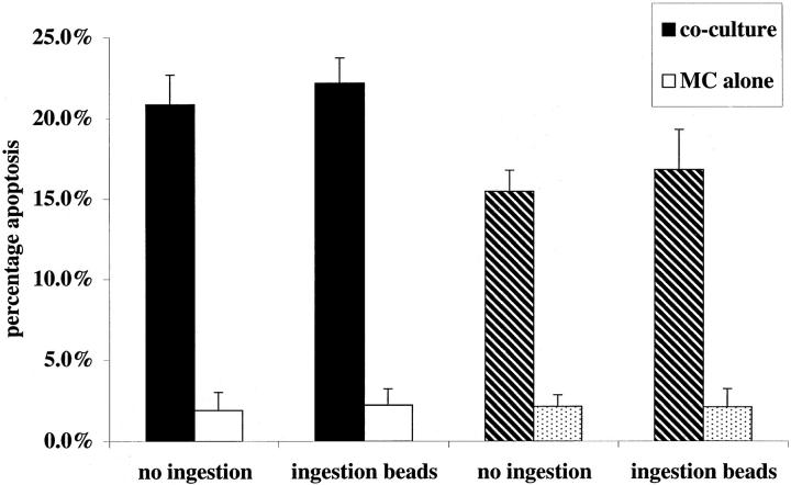
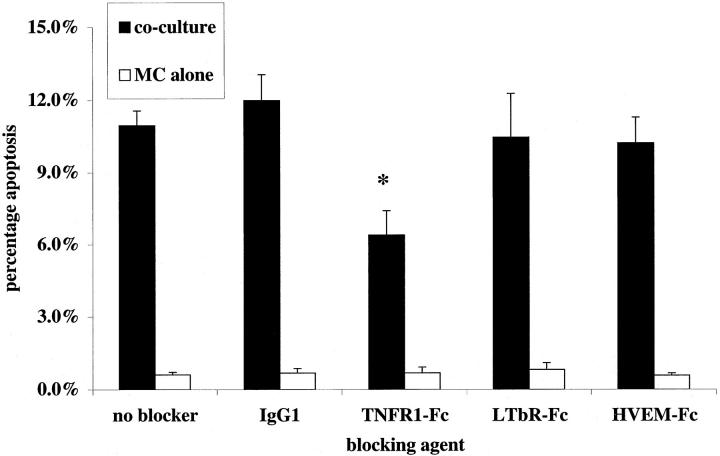
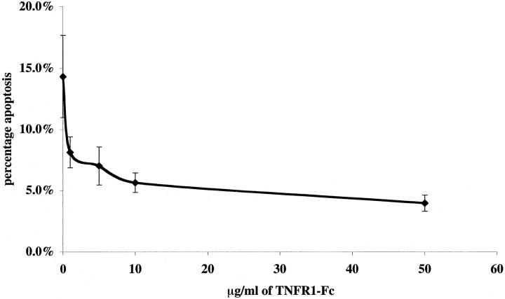
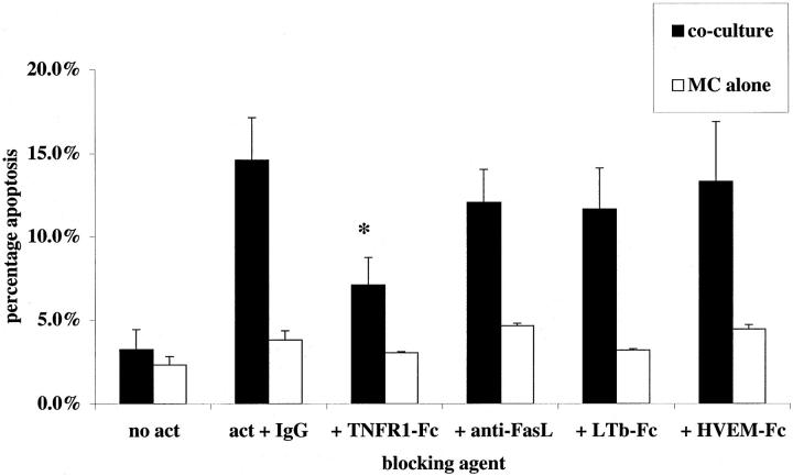
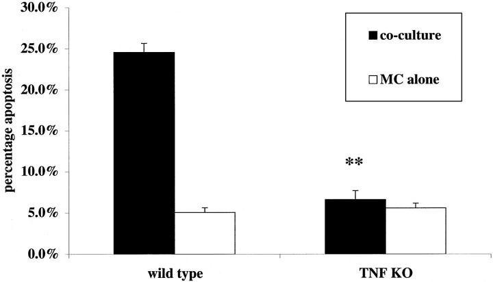
Activated macrophages (M(phi)) isolated from inflamed glomeruli or generated by interferon-gamma and lipopolysaccharide treatment in vitro induce glomerular mesangial cell apoptosis by hitherto incompletely understood mechanisms. In this report we demonstrate that nitric oxide-independent killing of co-cultured mesangial cells by interferon-gamma/lipopolysaccharide-activated M(phi) is suppressed by binding/ingestion of apoptotic cells and is mediated by tumor necrosis factor (TNF). Thus, soluble TNF receptor-1 significantly inhibited induction of mesangial cell apoptosis by 1) rodent M(phi) in the presence of nitric oxide synthase inhibitors or 2) human M(phi), both situations in which nitric oxide release was minimal. Furthermore, murine TNF knockout M(phi) were completely unable to induce mesangial cell apoptosis in the presence of nitric oxide synthase inhibitors. We conclude that TNF-restricted M(phi)-directed apoptosis of glomerular mesangial cells can be down-regulated by M(phi) binding/ingestion of apoptotic cells, suggesting a new mechanism for negative feedback regulation of M(phi) controls on resident cell number at inflamed sites.


Figures

Figure 1.
Figure 1.
Fluorescent micrograph (original magnification, ×320) showing activated co-culture of CMFDA green-labeled mesangial cells with Mφ. The co-culture was established according to Materials and Methods and activated with IFN-γ (100 U/ml) plus LPS (1 μg/ml) after 4 hours of exposure to preformed unlabeled apoptotic mesangial cells. At 24 hours, the co-culture was fixed and later counterstained with PI (1 μg/ml). Apoptotic green mesangial cells are clearly seen with red/orange pyknotic nuclear material and are readily distinguished from Mφ (arrows). Before fixation, co-culture was exposed to Hoechst 33342 (1 μg/ml) for confirmation of apoptosis of the green mesangial cells (see inset). Note inset field and arrow-marked apoptotic cells correspond to central field of main figure. Note also two apoptotic mesangial cells showing cytoplasmic blebbing (large arrows).
Figure 2.
Figure 2.
Induction of rodent mesangial cell apoptosis by activated Mφ is blocked by simultaneous phagocytosis of apoptotic mesangial cells when iNOS inhibitors are present. Established co-cultures of rodent Mφ and IFN-γ primed, CMFDA-labeled, mesangial cells, or mesangial cells growing alone (as control), were activated with IFN-γ (100 U/ml) and LPS (1 μg/ml). Simultaneously to activation, a fivefold excess of unlabeled apoptotic rodent mesangial cells was added (1 × 10 cells per well). In addition, to some wells (hatched bars), L-NMMA (200 μmol/L) was added at the same time as activation. After 24 hours of incubation, induction of CMFDA-labeled mesangial cells was scored in all wells. ***, P < 0.001 versus no ingestion (n = 5).
Figure 3.
Figure 3.
Induction of rodent mesangial cell apoptosis by activated Mφ is not blocked by simultaneous phagocytosis of latex beads when iNOS inhibitors are present. Both established co-cultures of rodent Mφ- and IFN-γ-primed, CMFDA-labeled, mesangial cells, and wells of mesangial cells alone were activated with IFN-γ (100 U/ml) and LPS (1 μg/ml). Simultaneously a fivefold excess of unlabeled sterile 10-μm latex beads was added (1 × 10 cells per well). In addition, to some wells (hatched bars), L-NMMA (200 μmol/L) was added. After 24 hours of incubation, induction of CMFDA-labeled mesangial cells was scored (n = 5).
Figure 4.
Figure 4.
Soluble TNFR1 inhibits nitric oxide-independent rodent Mφ killing of primed rodent mesangial cells. Murine bone marrow-derived Mφ were co-cultured with mesangial cells as described in Materials and Methods. Experiments were activated with IFN-γ and LPS as described, with L-NIL (30 μmol/L) to block NOS 2-mediated killing, and soluble Fc-fusion proteins TNFR1-Fc, LTβR-Fc, HVEM-Fc, or isotype control IgG1 were added in culture medium at 10 μg/ml. Control wells containing mesangial cells but no Mφ received all reagents. Mesangial cell apoptosis was assessed at 24 hours. Note a reduction in the capacity of Mφ to induce apoptosis in the presence of TNFR1-Fc. *, P < 0.05.
Figure 5.
Figure 5.
Concentration-dependent inhibition by sTNFR1 of nitric oxide independent rodent Mφ induction of rodent mesangial cell apoptosis. Using the same co-culture assay of mesangial cell apoptosis, a range of concentrations of TNFR1-Fc was added to wells and apoptosis quantified, compared with 50 μg/ml of IgG1 control shown as 0 μg/ml of TNFR1-Fc on the curve.
Figure 6.
Figure 6.
Induction of apoptosis of human mesangial cells by human monocyte-derived Mφ is mediated by TNFR1 ligation. Co-culture was established using primed human mesangial cells and Mφ. On activation with IFN-γ plus LPS, soluble Fc-fusion proteins TNFR1-Fc, LTβR-Fc, HVEM-Fc, or isotype control IgG1 (10 μg/ml) were added in culture medium at 10 μg/ml. Also, the blocking anti-Fas ligand antibody (1 μg/ml) was applied to adjacent wells. Control wells containing human mesangial cells but no Mφ received all reagents. Note a reduction in the capacity of Mφ to induce apoptosis in the presence of TNFR1-Fc, but not in the presence of any of the other reagents. *, P < 0.05.
Figure 7.
Figure 7.
The comparative effect of activated wild-type and Tnfα/Tnfβ−/− knockout murine Mφ on apoptosis of mesangial cells primed with IFN-γ. Mesangial cells were primed as described for 24 hours in the presence of IFN-γ (300 U/ml). Co-culture was established using bone marrow-derived Mφ from either Tnfα/Tnfβ−/− knockout mice or wild-type littermate controls and activated with IFN-γ (100 U/ml) plus LPS (1 μg/ml) in the presence of L-NIL (30 μmol/L) to block NOS 2-mediated killing. Experiments were assayed after 24 hours. Although the wild-type Mφ were able to induce mesangial cell apoptosis, the knockout animals incapable of inducing apoptosis greater than that seen in control wells. **, P < 0.01 compared with wild-type co-culture (n = 5).

References

    1. Schocklmann HO, Lang S, Sterzel RB: Regulation of mesangial cell proliferation. Kidney Int 1999, 56:1199-1207 - PubMed
    1. Baker AJ, Mooney A, Hughes J, Lombardi D, Johnson RJ, Savill J: Mesangial cell apoptosis: the major mechanism for resolution of glomerular hypercellularity in experimental mesangial proliferative nephritis. J Clin Invest 1994, 94:2105-2116 - PMC - PubMed
    1. Shimizu A, Masuda Y, Kitamura H, Ishizaki M, Sugisaki Y, Yamanaka N: Apoptosis in progressive crescentic glomerulonephritis. Lab Invest 1996, 74:941-951 - PubMed
    1. Duffield JS, Erwig LP, Wei X, Liew FY, Rees AJ, Savill JS: Activated macrophages direct apoptosis and suppress mitosis of mesangial cells. J Immunol 2000, 164:2110-2119 - PubMed
    1. Meszaros AJ, Reichner JS, Albina JE: Macrophage-induced neutrophil apoptosis. J Immunol 2000, 165:435-441 - PubMed

Publication types

Substances

LinkOut - more resources