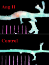
Angiotensin II increases urokinase-type plasminogen activator expression and induces aneurysm in the abdominal aorta of apolipoprotein E-deficient mice
- PMID: 11583973
- PMCID: PMC1850514
- DOI: 10.1016/S0002-9440(10)62532-1
Angiotensin II increases urokinase-type plasminogen activator expression and induces aneurysm in the abdominal aorta of apolipoprotein E-deficient mice
Abstract
Urokinase-type plasminogen activator (uPA) is increased in human abdominal aortic aneurysm (AAA). Chronic infusion of angiotensin II (Ang II) results in AAA in apolipoprotein E-deficient mice. We tested the hypothesis that Ang II infusion results in an elevation of uPA expression contributing to aneurysm formation. Ang II or vehicle was infused by osmotic pumps into apoE-KO mice. All mice treated with Ang II developed a localized expansion of the suprarenal aorta (75% increase in outer diameter), accompanied by an elevation of blood pressure (22 mmHg), compared to the vehicle-treated group. Histological examination of the dilated aortic segment revealed similarities to human AAA including focal elastin fragmentation, macrophage infiltration, and intravascular hemorrhage. Ang II treatment resulted in a 13-fold increase in the expression of uPA mRNA in the AAA segment in contrast to a twofold increase in the atherosclerotic aortic arch. Increased uPA protein was detected in the abdominal aorta as early as 10 days after Ang II infusion before significant aorta expansion. Thus, Ang II infusion results in macrophage infiltration, increased uPA activity, and aneurysm formation in the abdominal aorta of apoE-KO mice. These data are consistent with a causal role for uPA in the pathogenesis of AAA.
Figures


Similar articles
-
Urokinase-type plasminogen activator plays a critical role in angiotensin II-induced abdominal aortic aneurysm.Circ Res. 2003 Mar 21;92(5):510-7. doi: 10.1161/01.RES.0000061571.49375.E1. Epub 2003 Feb 13. Circ Res. 2003. PMID: 12600880
-
Effects of chymase inhibitor on angiotensin II-induced abdominal aortic aneurysm development in apolipoprotein E-deficient mice.Atherosclerosis. 2009 Jun;204(2):359-64. doi: 10.1016/j.atherosclerosis.2008.09.032. Epub 2008 Oct 8. Atherosclerosis. 2009. PMID: 18996524
-
Fasudil, a Rho-kinase inhibitor, attenuates angiotensin II-induced abdominal aortic aneurysm in apolipoprotein E-deficient mice by inhibiting apoptosis and proteolysis.Circulation. 2005 May 3;111(17):2219-26. doi: 10.1161/01.CIR.0000163544.17221.BE. Epub 2005 Apr 25. Circulation. 2005. PMID: 15851596
-
A peptide antagonist of thrombospondin-1 promotes abdominal aortic aneurysm progression in the angiotensin II-infused apolipoprotein-E-deficient mouse.Arterioscler Thromb Vasc Biol. 2015 Feb;35(2):389-98. doi: 10.1161/ATVBAHA.114.304732. Epub 2014 Dec 18. Arterioscler Thromb Vasc Biol. 2015. PMID: 25524772
-
Overexpression of PAI-1 prevents the development of abdominal aortic aneurysm in mice.Gene Ther. 2008 Feb;15(3):224-32. doi: 10.1038/sj.gt.3303069. Epub 2007 Nov 22. Gene Ther. 2008. PMID: 18033310
Cited by
-
Morphological and Biomechanical Differences in the Elastase and AngII apoE(-/-) Rodent Models of Abdominal Aortic Aneurysms.Biomed Res Int. 2015;2015:413189. doi: 10.1155/2015/413189. Epub 2015 May 3. Biomed Res Int. 2015. PMID: 26064906 Free PMC article.
-
Noninvasive imaging of vascular permeability to predict the risk of rupture in abdominal aortic aneurysms using an albumin-binding probe.Sci Rep. 2020 Feb 24;10(1):3231. doi: 10.1038/s41598-020-59842-2. Sci Rep. 2020. PMID: 32094414 Free PMC article.
-
Total lymphocyte deficiency attenuates AngII-induced atherosclerosis in males but not abdominal aortic aneurysms in apoE deficient mice.Atherosclerosis. 2010 Aug;211(2):399-403. doi: 10.1016/j.atherosclerosis.2010.02.034. Epub 2010 Mar 4. Atherosclerosis. 2010. PMID: 20362292 Free PMC article.
-
Monocytes and macrophages in abdominal aortic aneurysm.Nat Rev Cardiol. 2017 Aug;14(8):457-471. doi: 10.1038/nrcardio.2017.52. Epub 2017 Apr 13. Nat Rev Cardiol. 2017. PMID: 28406184 Review.
-
Weight loss in obese C57BL/6 mice limits adventitial expansion of established angiotensin II-induced abdominal aortic aneurysms.Am J Physiol Heart Circ Physiol. 2010 Jun;298(6):H1932-8. doi: 10.1152/ajpheart.00961.2009. Epub 2010 Mar 19. Am J Physiol Heart Circ Physiol. 2010. PMID: 20304811 Free PMC article.
References
-
- Freestone T, Turner RJ, Coady A, Higman DJ, Greenhalgh RM, Powell JT: Inflammation and matrix metalloproteinases in the enlarging abdominal aortic aneurysm. Arterioscler Thromb Vasc Biol 1995, 15:1145-1151 - PubMed
-
- Davis V, Persidskaia R, Baca-Regen L, Itoh Y, Nagase H, Persidsky Y, Ghorpade A, Baxter BT: Matrix metalloproteinase-2 production and its binding to the matrix are increased in abdominal aortic aneurysms. Arterioscler Thromb Vasc Biol 1998, 18:1625-1633 - PubMed
-
- Thompson RW, Holmes DR, Mertens RA, Liao S, Botney MD, Mecham RP, Welgus HG, Parks WC: Production and localization of 92-kilodalton gelatinase in abdominal aortic aneurysms. An elastolytic metalloproteinase expressed by aneurysm-infiltrating macrophages. J Clin Invest 1995, 96:318-326 - PMC - PubMed
MeSH terms
Substances
Grants and funding
LinkOut - more resources
Full Text Sources
Molecular Biology Databases
Research Materials
Miscellaneous

