. 2001 Oct;159(4):1465-75.
doi: 10.1016/S0002-9440(10)62533-3.

Dissection of key events in tubular epithelial to myofibroblast transition and its implications in renal interstitial fibrosis

Affiliations

Dissection of key events in tubular epithelial to myofibroblast transition and its implications in renal interstitial fibrosis

J Yang et al. Am J Pathol. 2001 Oct.

Abstract

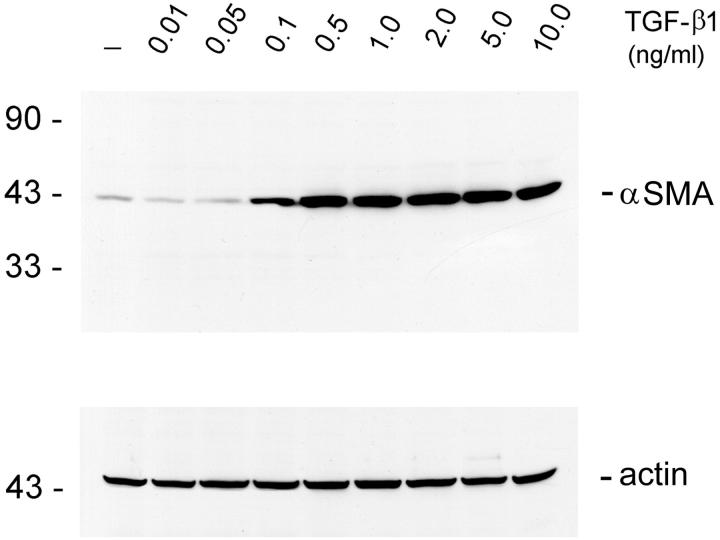
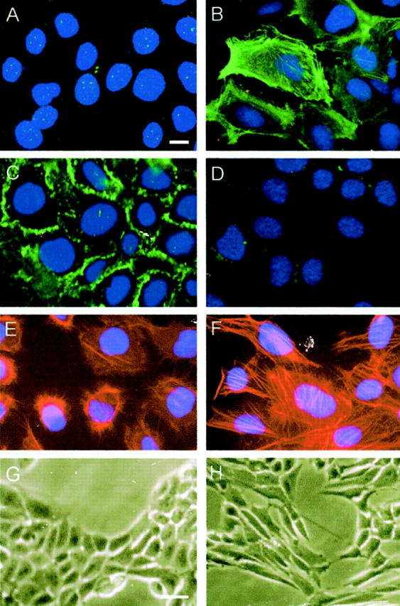
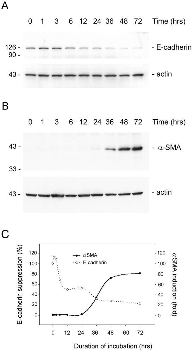
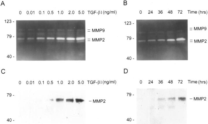
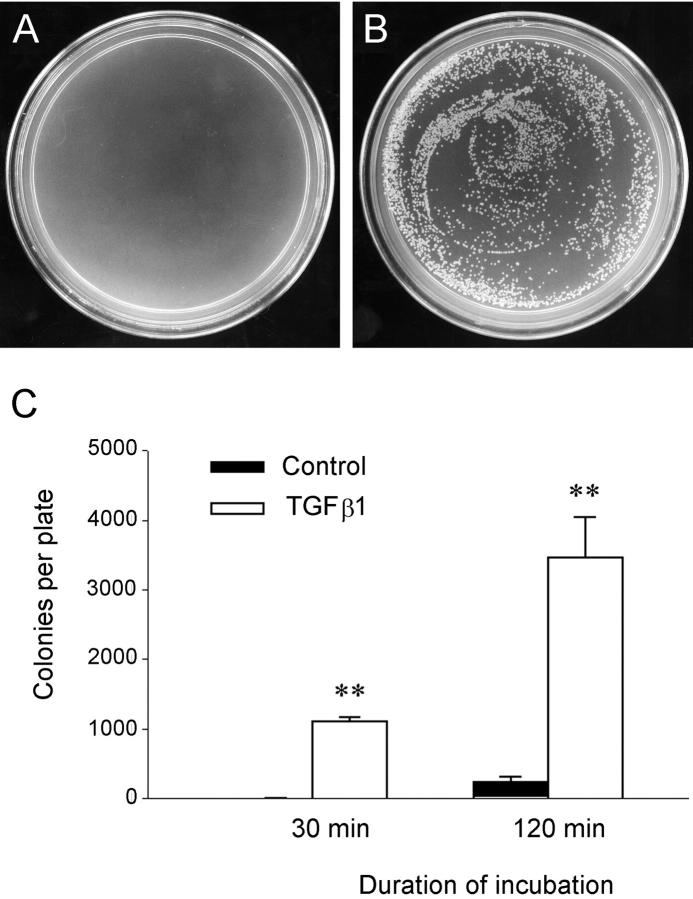
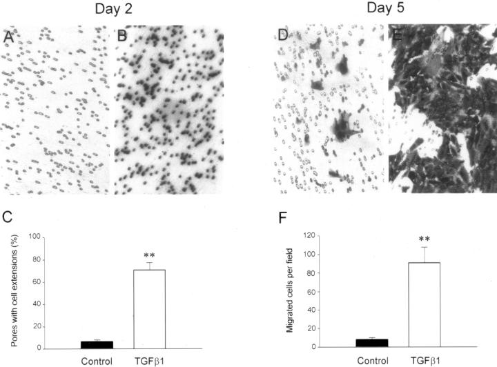
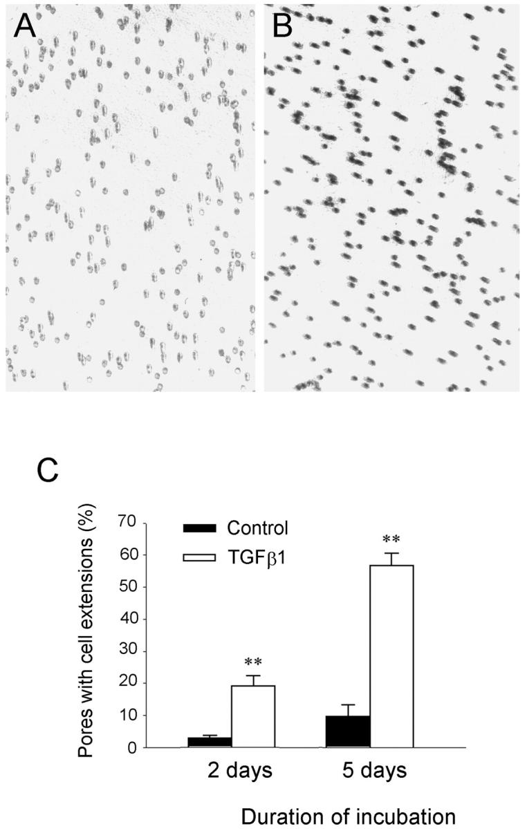
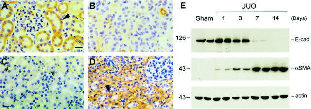
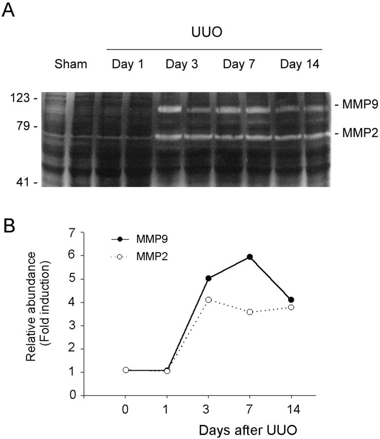
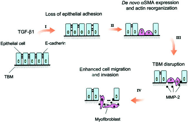
Myofibroblast activation is a key event playing a critical role in the progression of chronic renal disease. Emerging evidence suggests that myofibroblasts can derive from tubular epithelial cells by an epithelial to mesenchymal transition (EMT); however, the details regarding the conversion between these two cell types are poorly understood. Here we dissect the key events during the process of EMT induced by transforming growth factor-beta1. Incubation of human tubular epithelial cells with transforming growth factor-beta1 induced de novo expression of alpha-smooth muscle actin, loss of epithelial marker E-cadherin, transformation of myofibroblastic morphology, and production of interstitial matrix. Time-course studies revealed that loss of E-cadherin was an early event that preceded other alterations during EMT. The transformed cells secreted a large amount of matrix metalloproteinase-2 that specifically degraded tubular basement membrane. They also exhibited an enhanced motility and invasive capacity. These alterations in epithelial phenotypes in vitro were essentially recapitulated in a mouse model of renal fibrosis induced by unilateral ureteral obstruction. Hence, these results indicate that tubular epithelial to myofibroblast transition is an orchestrated, highly regulated process involving four key steps including: 1) loss of epithelial cell adhesion, 2) de novo alpha-smooth muscle actin expression and actin reorganization, 3) disruption of tubular basement membrane, and 4) enhanced cell migration and invasion.


Figures

Figure 1.
Figure 1.
TGF-β1 induces de novo expression of α-SMA in tubular epithelial cells. HKC cells treated without (control) or with different amounts of TGF-β1 for 72 hours in serum-free medium. The whole cell lysate was immunoblotted with a specific α-SMA antibody. The same blot was reprobed with β-actin to ensure equal loading of each lane.
Figure 2.
Figure 2.
Tubular epithelial to myofibroblast transition in vitro. HKC cells treated without (left column) or with 4 ng/ml of TGF-β1 (right column) for 72 hours in serum-free medium. The myofibroblast or epithelial cell markers were detected by an indirect immunostaining. The transformed cells acquired α-SMA (A, B), lost E-cadherin (C, D), formed stress fiber by actin reorganization (E, F), and displayed myofibroblast morphology (G, H). Scale bars: 10 μm (A–F), 20 μm (G and H).
Figure 3.
Figure 3.
Loss of E-cadherin is an early event during epithelial to myofibroblast transition. HKC cells were treated with TGF-β1 for various periods of time as indicated in serum-free medium. The whole cell lysate was immunoblotted with antibodies against α-SMA, E-cadherin, and β-actin, respectively. A: Time-course of E-cadherin expression. B: Time-dependency of α-SMA expression. C: Graphic presentation of the relative abundance of E-cadherin and α-SMA after TGF-β1-induced cell phenotypic transition.
Figure 4.
Figure 4.
TGF-β1 induces MMP expression in tubular epithelial cells. A and B: Zymographic analysis of the conditioned media derived from HKC cells treated without (control) or with different amounts of TGF-β1 for various periods of time as indicated. Samples equalized for protein content were separated on a 10% SDS-polyacrylamide gel containing 0.1% gelatin. Proteolytic activity is demonstrated by digestion of gelatin, resulting in the bands of clearing. The locations of bands corresponding to pro- and active MMP-2 as well as pro- and active MMP-9 are indicated. C and D: Western blot analyses of the conditioned media from HKC cells treated without (control) or with various concentration of TGF-β1 for different periods of time in the serum-free medium. The location of a 72-kd band corresponding to MMP-2 is indicated. A and C: HKC cells were incubated with different concentrations of TGF-β1 as indicated for 72 hours. B and D: HKC cells were incubated with 2 ng/ml of TGF-β1 for various periods of time as indicated.
Figure 5.
Figure 5.
Functional disruption of the reconstituted TBM by the conditioned media from the transformed cells. Matrigels (15-μm depth) analogous to native TBM matrix were formed on the transwell filters and incubated with the conditioned media from either control or TGF-β1-treated HKC cells for 4 days. The integrity of Matrigels was assessed by bacterial translocation through the gels. A and B: Representative plates show the colonies formed from the bacteria passed through the Matrigels. C: Graphic presentation of the numbers of bacteria translocated via the gels. **, P < 0.01 versus control (n = 3).
Figure 6.
Figure 6.
Enhanced migration of the transformed cells. HKC cells were seeded on the transwell filters of Boyden chamber (pore size, 8 μm) and incubated with or without TGF-β1 for 2 and 5 days, respectively. The cells or cell extensions that passed through the pores of filters were counted after staining. A–C: Most pores of the transwell filters were filled with cell extensions after incubation with TGF-β1 for 2 days, but no cells migrated through the pores to the opposite side of filters. D–F: After incubation with TGF-β1 for 5 days, the transformed cells migrated through the pores to the opposite side of filters. A, B, D, E: Representative micrographs of the transwell filters. C and F: Graphic presentation of the numbers of cells or cell extensions migrated through the pores of the filters after incubation with or without TGF-β1 for 2 and 5 days, respectively. **, P < 0.01 versus control (n = 3).
Figure 7.
Figure 7.
Increased invasive capacity of the transformed cells on Matrigel. HKC cells were seeded on the top of Matrigels in the transwell filters of the Boyden chamber and incubated with or without TGF-β1 for 2 and 5 days, respectively. The cell extensions that migrated across the Matrigels and passed through the pores of filters were counted after staining. A and B: Representative micrographs show the pores of the transwell filters filled with cell extensions after incubation without or with TGF-β1 for 5 days. C: Graphic presentation of the numbers of pores filled with cell extensions after incubation with or without TGF-β1 for 2 and 5 days, respectively. **, P < 0.01 versus control (n = 3).
Figure 8.
Figure 8.
Expression of both TGF-β1 and its type I receptor increases rapidly and specifically in renal tubular epithelia in mouse model of renal interstitial fibrosis induced by UUO. A: Western blot demonstrates an early induction of TGF-β type I receptor expression in the obstructed kidneys after UUO. Representative picture shows the immunoblotting results of two animals per time point. Marked TβR-I elevation was observed as early as 1 day after UUO. B: Quantitative determination of TGF-β1 protein by enzyme-linked immunosorbent assay reveals a time-dependent induction of TGF-β1 in the obstructed kidneys. Data are presented as mean ± SE from four animals per group. *, P < 0.05; **, P < 0.01 versus sham. Representative micrographs show the localization of TGF-β type I receptor (C and D) and TGF-β1 (E and F) in the kidneys at 7 days after UUO (D and F) or sham operation (C and E). Both TGF-β1 and TβR-I are specifically induced in renal tubular epithelia after UUO (F and D). Asterisk indicates positively stained tubules for both TGF-β1 and TβR-I in serial sections. Scale bar, 20 μm.
Figure 9.
Figure 9.
Alterations of protein expression pattern in the obstructed kidneys recapitulate key events in tubular epithelial to myofibroblast transition. Kidney sections were prepared from mice at 7 days after either UUO or sham operation and stained with various specific antibodies. Representative micrographs show the alterations in protein expression pattern after UUO. A and C: Sham-operated mice. B and D: UUO mice. A and B: E-cadherin. C and D: α-SMA. Scale bar, 20 μm. Arrowheads indicate positively stained cells. E: Western blot demonstrates a time-dependent alteration in the expression of E-cadherin and α-SMA in the obstructed kidneys after UUO. Representative pictures show the results of two animals per time point.
Figure 10.
Figure 10.
Induction of renal MMP-2 and -9 expressions in vivo. Kidney tissues were collected from mice receiving UUO at different time points as indicated. A: Zymographic analysis of whole kidney lysates indicates that renal MMP-2 and -9 expressions were markedly induced in a time-dependent manner. B: Graphic presentation of the relative abundance (fold induction) of MMP-2 and MMP-9 in the kidneys at different time points after UUO.
Figure 11.
Figure 11.
Schematic illustration depicting four key events during tubular epithelial to myofibroblast transition. The diagram illustrates the four key events essential for the completion of entire EMT course at cellular level, which include: 1) loss of epithelial adhesion properties, 2) de novo expression of α-SMA and actin reorganization, 3) disruption of the TBM, and 4) enhanced migration and invasive capacity of the transformed cells. TGF-β1 as a sole factor is capable of inducing tubular epithelial cells to undergo all four steps.

References

    1. Port FK, Fenton SSA, Mazzuchi N: ESRD throughout the world: morbidity, mortality and quality of life. Kidney Int 2000, 57:S1-S2
    1. Eddy AA: Molecular insights into renal interstitial fibrosis. J Am Soc Nephrol 1996, 7:2495-2508 - PubMed
    1. Remuzzi G, Bertani T: Pathophysiology of progressive nephropathies. N Engl J Med 1998, 339:1448-1456 - PubMed
    1. Roberts IS, Burrows C, Shanks JH, Venning M, McWilliam LJ: Interstitial myofibroblasts: predictors of progression in membranous nephropathy. J Clin Pathol 1997, 50:123-127 - PMC - PubMed
    1. Essawy M, Soylemezoglu O, Muchaneta-Kubara EC, Shortland J, Brown CB, el Nahas AM: Myofibroblasts and the progression of diabetic nephropathy. Nephrol Dial Transplant 1997, 12:43-50 - PubMed

Publication types

MeSH terms