. 2001 Oct;159(4):1477-94.
doi: 10.1016/S0002-9440(10)62534-5.

Mitochondrial alterations caused by defective peroxisomal biogenesis in a mouse model for Zellweger syndrome (PEX5 knockout mouse)

Affiliations

Mitochondrial alterations caused by defective peroxisomal biogenesis in a mouse model for Zellweger syndrome (PEX5 knockout mouse)

E Baumgart et al. Am J Pathol. 2001 Oct.

Abstract

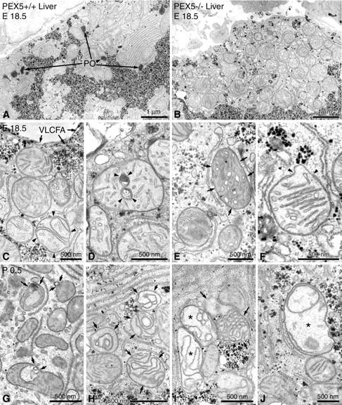
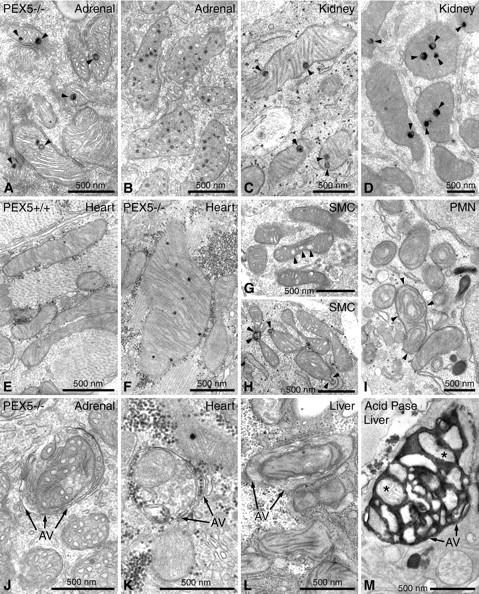
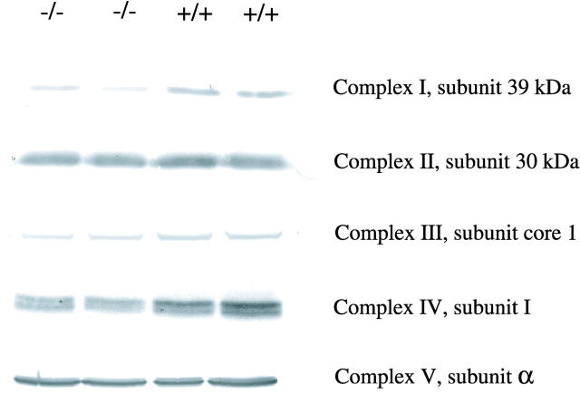
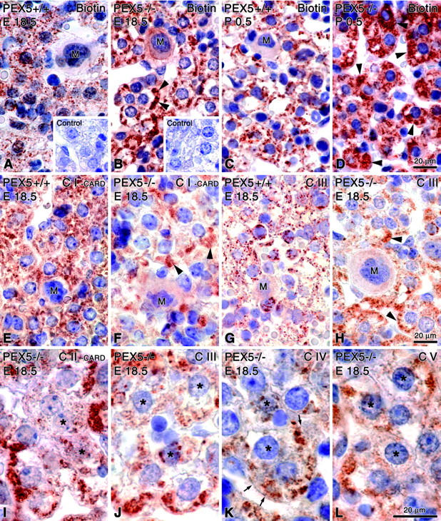
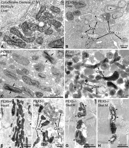
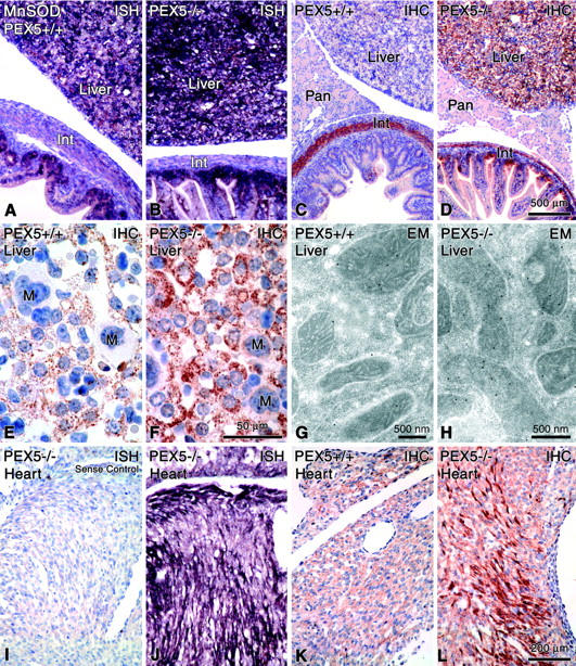
Zellweger syndrome (cerebro-hepato-renal syndrome) is the most severe form of the peroxisomal biogenesis disorders leading to early death of the affected children. To study the pathogenetic mechanisms causing organ dysfunctions in Zellweger syndrome, we have recently developed a knockout-mouse model by disrupting the PEX5 gene, encoding the targeting receptor for most peroxisomal matrix proteins (M Baes, P Gressens, E Baumgart, P Carmeliet, M Casteels, M Fransen, P Evrard, D Fahimi, PE Declercq, D Collen, PP van Veldhoven, GP Mannaerts: A mouse model for Zellweger syndrome. Nat Genet 1997, 17:49-57). In this study, we present evidence that the absence of functional peroxisomes, causing a general defect in peroxisomal metabolism, leads to proliferation of pleomorphic mitochondria with severe alterations of the mitochondrial ultrastructure, changes in the expression and activities of mitochondrial respiratory chain complexes, and an increase in the heterogeneity of the mitochondrial compartment in various organs and specific cell types (eg, liver, proximal tubules of the kidney, adrenal cortex, heart, skeletal and smooth muscle cells, neutrophils). The changes of mitochondrial respiratory chain enzymes are accompanied by a marked increase of mitochondrial manganese-superoxide dismutase, as revealed by in situ hybridization and immunocytochemistry, suggesting increased production of reactive oxygen species in altered mitochondria. This increased oxidative stress induced probably by defective peroxisomal antioxidant mechanisms combined with accumulation of lipid intermediates of peroxisomal beta-oxidation system could contribute significantly to the pathogenesis of multiple organ dysfunctions in Zellweger syndrome.


Figures

Figure 1.
Figure 1.
Electron micrographs of liver sections stained with alkaline DAB for the localization of peroxisomal catalase activity and postfixed with reduced osmium. A: Low-magnification view of an hepatocyte from a control animal of gestational age E18.5. Peroxisomes (PO) are easily recognized by the DAB reaction product in their matrix and are often found in the vicinity of glycogen areas. B: Low-magnification view of a similar region of an hepatocyte in a liver section of a PEX5−/− mouse. Note the absence of peroxisomes near the glycogen areas and the strong proliferation of pleomorphic mostly round, large mitochondria, some of which exhibit stacks of parallel cristae. C–F: Higher magnification views of altered mitochondria in hepatocytes of PEX5−/− mice (E18.5). C: Curvilinear alterations of cristae are already observed in some hepatic mitochondria of PEX5−/− mice before birth (arrowheads). Because of the defect in peroxisomal β-oxidation, crystals of very long chain fatty acids (VLCFA) accumulate on the surface of regular lipid droplets (arrows). D: Myelin-like or ring-shaped alterations of mitochondrial cristae (arrowheads). E: Mitochondria with tubular cristae (small arrows). F: A mitochondrion with parallel-stacked cristae, exhibiting a focal disruption of the outer membrane (arrowheads) and protrusion of the inner membrane through the gap. G–J: Mitochondrial alterations in hepatocytes of newborn PEX5−/− animals (P0.5) that are more severe than in E18.5. G: Mitochondria with evaginations and invaginations of the outer membrane (small arrows). H: Severe curvilinear alterations of mitochondrial cristae (small arrows), with the formation of different types of mitochondrial ghosts (I, small arrows). I and J: Swollen mitochondria with less dense matrix and curvilinear cristae (I, asterisks) or rarefication of cristae (J, asterisk).
Figure 2.
Figure 2.
Mitochondrial alterations of extrahepatic cells of PEX5−/− mice of gestational stage E18.5, stained for the localization of peroxisomal catalase activity with the alkaline DAB method (A–L) or for the localization of lysosomal acid-phosphatase activity with cerium chloride (M). A and B: Mitochondria in epithelial cells of the zona fasciculata of the adrenal gland with large lamellated granules (arrowheads) (A) and accumulation of electron-dense material in the intermembrane space of cristae (B). C and D: Kidney, large inclusions in mitochondrial matrix of proximal tubules (arrowheads). E and F: Cardiomyocytes of normal (E) and PEX5−/−(F) mice. Megamitochondria with an increase in the number of dense granules are shown (F) as compared to the low number of granules and regular size of mitochondria (E) in a control animal of the same gestational stage (E18.5). G and H: Smooth muscle cells (SMCs) of the intestinal wall of PEX5−/− mice showing enlargement of the intermembrane space in cristae (arrowheads, G) and in- or evaginations of the mitochondrial outer membrane (arrowheads, H). I: A neutrophil (PMN) with several abnormal mitochondria with circular cristae (arrowheads), in a focus of extramedullary hematopoiesis of a PEX5−/− mouse liver. J–L: Autophagic vacuoles (AVs) containing mitochondria in an epithelial cell of the zona fasciculata of the adrenal gland (J), a cardiomyocyte (K), or a hepatocyte (L). M: By using the cytochemical staining technique for acid phosphatase activity with cerium chloride, large autophagic vacuoles containing many mitochondria (two of them marked with asterisks) are identified in a hepatocyte of a PEX5−/− mouse.
Figure 3.
Figure 3.
Western blots of subunits of the respiratory chain complexes in liver homogenates of newborn (P0.5) PEX5-deficient and control mice. Thirty μg of total protein was loaded onto an SDS-PAGE gel under reducing conditions, except for complex I (15 μg). The blots were incubated with antibodies against the 39-kd subunit of complex I, the 30-kd subunit of complex II, the subunit core 1 of complex III, subunit 1 of complex IV, and the α subunit of complex V. Note the reductions of complexes I, III, and IV.
Figure 4.
Figure 4.
Light microscopical visualization of mitochondria with different techniques in paraffin sections of the liver of PEX5+/+ (E18.5: A, E, G; P0.5: C) or PEX5−/− (E18.5: B, F, H, I–L; P0.5: D) mice. A–D: Localization of biotinylated proteins in mitochondria with extravidin-peroxidase. Insets in A and B depict appropriate negative controls after blocking of endogenous biotin groups. Note the granular appearance of mitochondria in control mice (A and C) in contrast to the large subsinusoidal aggregates (arrowheads) in PEX5-deficient E18.5 mice (B). In P0.5 animals the mitochondrial aggregates fill up the cytoplasm of most PEX5−/− hepatocytes (D, arrowheads). E–L: Indirect immunohistochemical localization of distinct proteins of different mitochondrial respiratory chain complexes (CI to CV) with appropriate antibodies. For optimal visualization, the signals for complexes I and II had to be amplified with catalyzed reporter deposition (E, F, I). Hepatocytes in the liver of PEX5−/− mice with strong proliferation of mitochondria are indicated by arrowheads (F and H). Asterisks in I–L indicate hepatocytes with heterogeneous staining of their mitochondrial populations (marked in detail with small arrows in K). M, megakaryocytes. Note the large aggregates of proliferated mitochondria in hepatocytes of PEX5−/− mice. Bars on the right indicate magnifications for all pictures in the corresponding row.
Figure 5.
Figure 5.
Enzyme cytochemical staining for cytochrome c oxidase (Cox) activity in mitochondria of hepatocytes (A–C), of cardiomyocytes (D–F), and of myocytes of the diaphragm (Skel. M., skeletal muscle) (G, H) in control (A, E) and in PEX5−/− mice (B–D, F–H) of gestational age E18.5. Note the marked intracellular heterogeneity of Cox activity in hepatocytes (B, C) and cardiomyocytes (D, F) of PEX5−/− mice. B: A swollen mitochondrion with circular cristae and a negative Cox reaction is marked with an asterisk; mitochondria with a fluffy matrix and negative Cox reaction (Cox−) are marked with arrowheads. C: Note the strong Cox activity in mitochondria with parallel-stacked cristae in hepatocytes of PEX5−/− mice (small arrows). G, H: Mitochondria in myocytes of the diaphragm with altered, curvilinear cristae and positive Cox activity are marked with arrowheads.
Figure 6.
Figure 6.
Detection of MnSOD mRNA (A, B, J; negative control = I) by nonradioactive in situ hybridization, MnSOD protein (C-F, K, L) by immunohistochemistry (IHC), and G, H by immunoelectron microscopy (EM) in different organs of control (A, C, E, G, K) and PEX5−/− mice (B, D, F, H, I, J, L). Note the strong increases of MnSOD mRNA and protein in the liver (B, D, F) and the heterogeneous induction of mRNA and proteins in cardiomyocytes (J, L). Proliferated mitochondria in hepatocytes of PEX5−/− mice are stronger stained for MnSOD protein (F) than the mitochondria of control animals (E) and by electron microscopy the immunolabeling is significantly higher in PEX5-deficient mice (H) than in controls (G). Int, intestine; Pan, pancreas; M, megakaryocytes. Bars on the right indicate magnifications for all figures in the corresponding row (except for electron micrographs).

References

    1. Baes M, Gressens P, Baumgart E, Carmeliet P, Casteels M, Fransen M, Evrard P, Fahimi D, Declercq PE, Collen D, van Veldhoven PP, Mannaerts GP: A mouse model for Zellweger syndrome. Nat Genet 1997, 17:49-57 - PubMed
    1. De Duve C, Baudhuin P: Peroxisomes (microbodies and related particles). Physiol Rev 1966, 46:323-357 - PubMed
    1. Singh I: Mammalian peroxisomes: metabolism of oxygen and reactive oxygen species. Ann NY Acad Sci 1996, 804:612-627 - PubMed
    1. Lopez-Huertas E, Charlton WL, Johnson B, Graham IA, Baker A: Stress induces peroxisome biogenesis genes. EMBO J 2000, 19:6770-6777 - PMC - PubMed
    1. Schrader M, Wodopia R, Fahimi HD: Induction of tubular peroxisomes by UV irradiation and reactive oxygen species in HepG2 cells. J Histochem Cytochem 1999, 47:1141-1148 - PubMed

Publication types

MeSH terms

Substances