. 2001 Oct;159(4):1555-66.
doi: 10.1016/S0002-9440(10)62540-0.

Matrix metalloproteinases: a role in the contraction of vitreo-retinal scar tissue

Affiliations

Matrix metalloproteinases: a role in the contraction of vitreo-retinal scar tissue

C M Sheridan et al. Am J Pathol. 2001 Oct.

Abstract

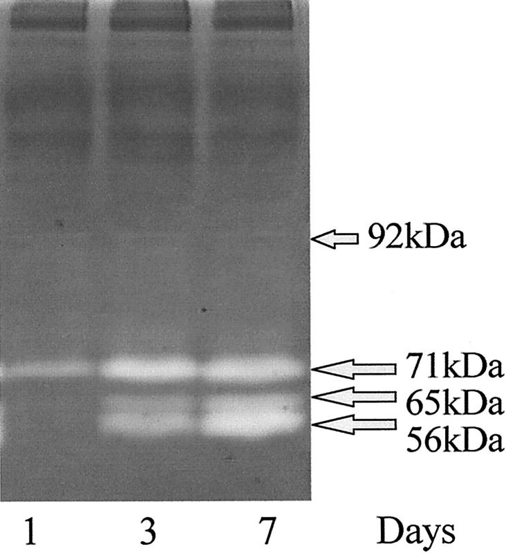
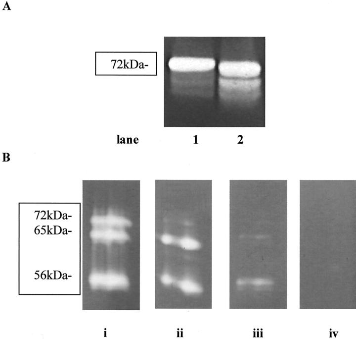
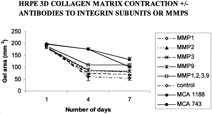
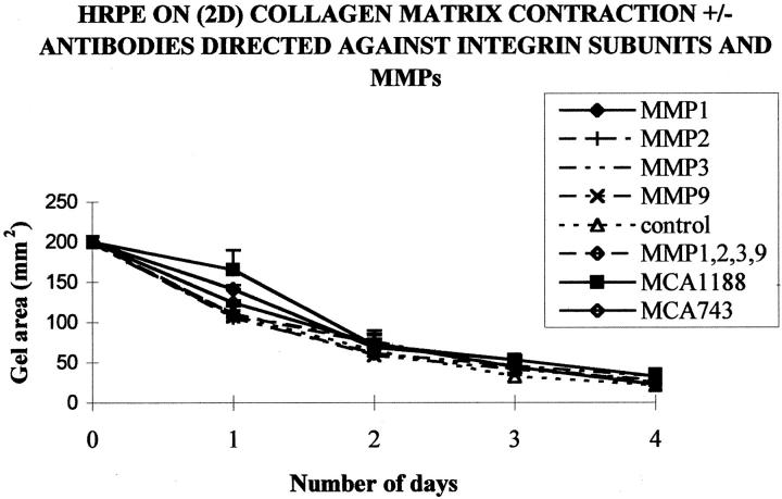
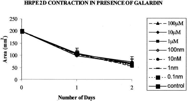
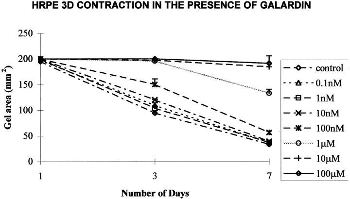
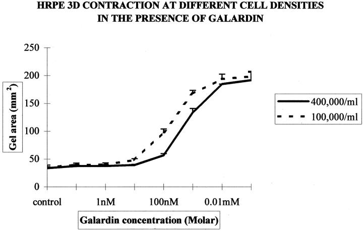
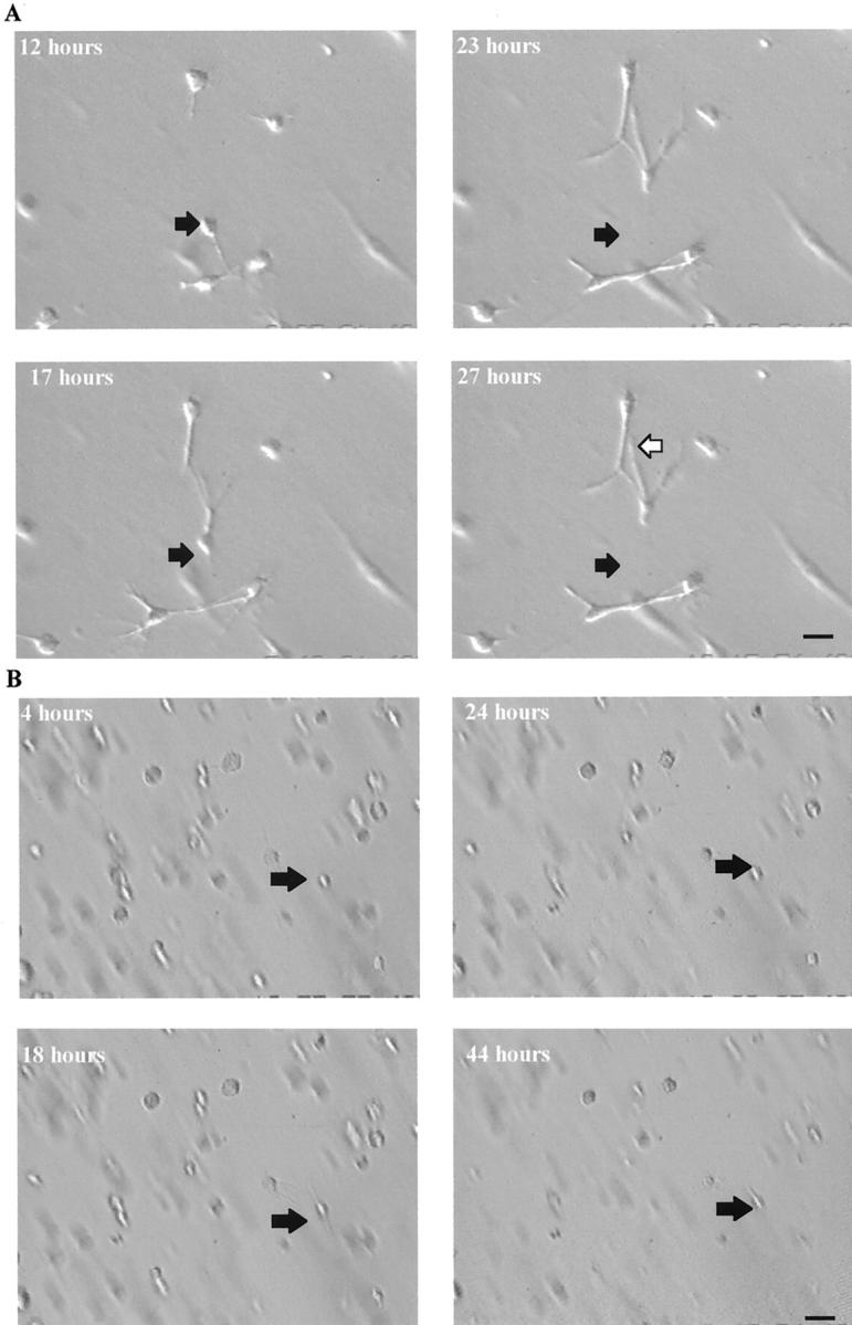
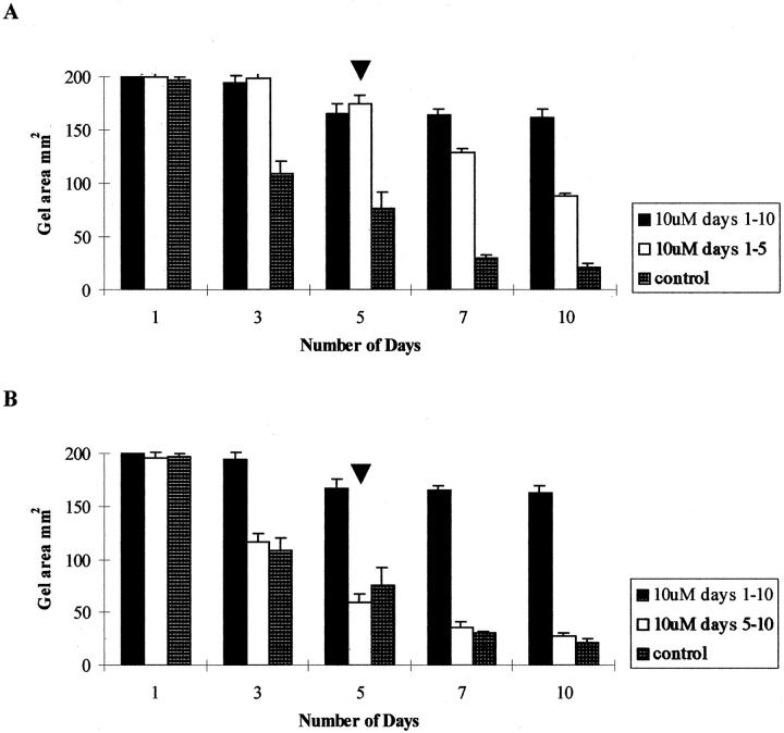
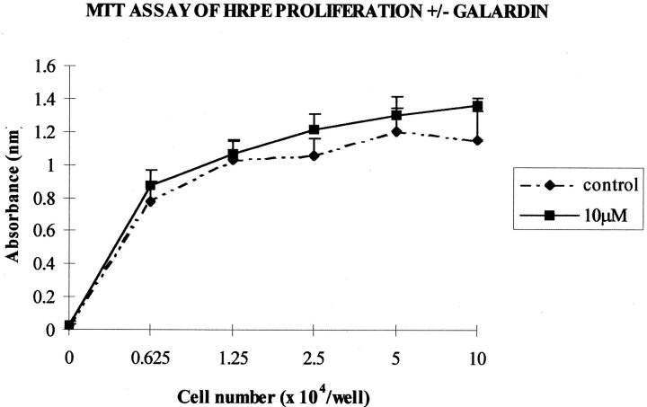
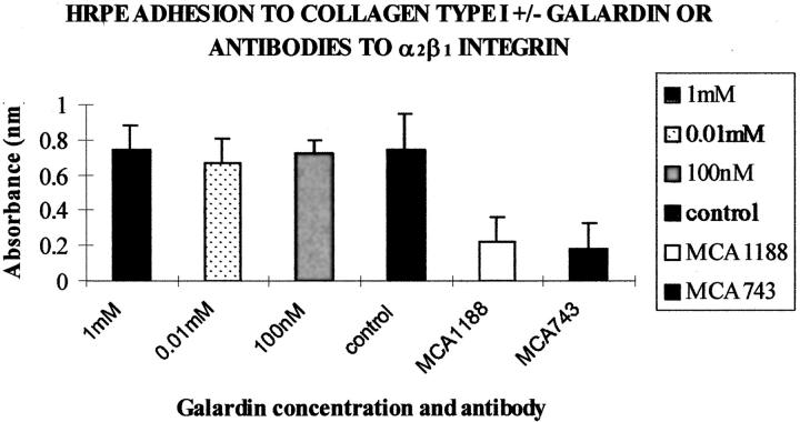
The most common cause of failure of retinal reattachment surgery is formation of fibrocellular contractile membranes on both surfaces of the neuroretina. This intraocular fibrosis, known as proliferative vitreoretinopathy, results in a blinding tractional retinal detachment because of the contractile nature of the membrane. Contractility is a cell-mediated event that is thought to be dependent on locomotion and adhesion to the extracellular matrix. Interactions between cells and the extracellular matrix can be influenced by matrix metalloproteinases (MMPs) and we investigated the role of MMPs in two in vitro models (two- and three-dimensional) of human retinal pigment epithelial (RPE) cell-mediated contraction. MMP activity was detected using enzyme-linked immunosorbent assays and zymography techniques that revealed MMP-1, -2, -3, and -9 positivity during the collagen matrix contraction assays. RPE-populated collagen matrix contraction (three-dimensional) was inhibited using a cocktail of anti-MMP antibodies and with Galardin (a broad-spectrum MMP inhibitor). Galardin inhibition was dose-dependent, reversible, and dependent on cell number. MMP inhibitors had no effect on contraction when RPEs were seeded on two-dimensional collagen matrices or on cellular adhesion to collagen type I. Our results suggest that MMP activity may be required for three-dimensional but not two-dimensional RPE-collagen matrix contraction.
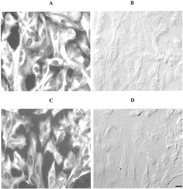

Figures

Figure 1.
Figure 1.
A zymogram gel in which media collected from contracting three-dimensional collagen matrices at time points of days 1, 3, and 7 after seeding. A similar profile was observed for both cells on and in the matrices. The molecular weights of the major bands indicated the presence of MMP-2 in both its latent (71 kd) and active (65 kd) form and MMP-1 or MMP-3 at 56 kd. All three bands were seen to increase in intensity during of contraction of the gels. Two very faint bands could just be seen at similar intensities on all days studied at molecular weights corresponding to the presence of MMP-9 at 100 and 92 kd.
Figure 2.
Figure 2.
A: A zymogram in which samples have been run after incubation with (lane 2) and without (lane 1) APMA. After activation with APMA there is a small reduction of the proenzyme (arrow) at ∼ 72 kd and an increase in the smaller active 65-kd band. B: Zymograms in which samples of media (day 7) were incubated with increasing concentrations of phenanthroline within the developing buffer. A reduction in the amount of gelatinolytic activity produced during collagen gel contraction is observed with increasing concentrations of phenanthroline (i = developing buffer only, ii (200 nmol/L), iii (200 μm), and iv (20 mmol/L) show the final concentrations of phenanthroline within the developing buffer).
Figure 3.
Figure 3.
Graph representing the mean collagen matrix area (±SD) for HRPE-populated collagen matrices (three-dimensional) either in the presence or absence of individual antibodies directed against either β-1 integrin (MCA1188); α-2 integrin (MCA743); MMP-1, -2, -3, and -9; or a cocktail of antibodies directed against all four of the above MMPs. A significant (P < 0.001) inhibition of contraction was observed in matrices incubated with antibody (10 μg/ml) directed against both the integrin subunits on day 4 and the MCA743 antibody maintained this inhibition on the seventh day. At no time point was there any significant difference between single MMP antibody-treated and control groups (P > 0.05). A significant inhibition of contraction occurred on days 4 and 7 with the cocktail of antibodies (P < 0.05).
Figure 4.
Figure 4.
Graph showing the mean collagen matrix area for HRPE seeded on two-dimensional collagen matrices either in the presence or absence of antibodies directed against MMP-1, -2, -3, or -9. At no time point was there a significant difference (P > 0.05) between control and any MMP antibody-treated groups. Significant inhibition of contraction occurred at day 1 in matrices containing the antibody (10 μg/ml) directed against the β-1 integrin subunit (MCA 1188; P < 0.05; n = 6).
Figure 5.
Figure 5.
Graph illustrates HRPE cell-mediated contraction for cells seeded on collagen matrices (two-dimensional) in the presence of MMP inhibitor Galardin. The presence of Galardin did not significantly alter the rate or extent of matrix contraction at any concentration. Data are represented as means ±SD.
Figure 6.
Figure 6.
Graph illustrating the mean gel area (SD) against time for HRPE-populated collagen gels (three-dimensional) surrounded with media containing different concentrations of Galardin. From day 3 onwards, 10 nmol/L of Galardin and above significantly (P < 0.05) inhibited collagen gel contraction compared to controls.
Figure 7.
Figure 7.
Graph showing the mean gel area (mm2) at day 7 for HRPE-populated matrices containing either 4 × 10 or 1 × 10 cells/ml that have undergone contraction with different concentrations of Galardin (0.1 nmol/L to 100 μmol/L). High-populated matrices require a greater concentration of inhibitor to exert anti-contractile effects. The inhibition of contraction appears in a cell number- and dose-dependant manner.
Figure 8.
Figure 8.
Photomicrographs of whole matrices fluorescently immunostained for CK18 (A–D). During collagen matrix contraction the cells were initially round with small processes extending within the matrix (A; day 1) and were later seen to adopt a stellate and spindle-shaped appearance by day 7 (B). The cells in matrices incubated with Galardin only extended out small processes into the surrounding matrix (C, arrows; day 1) whereas in control cell-populated matrices, the cells characteristically were stellate in appearance and even formed tunnels within the matrix (D, arrowheads; scale bars, 10 μm). Electron micrographs of matrices (E and F) illustrating that no holes or cells are present on the matrix surface, early in the contractile process (day 1; F). RPE cells can still be visualized just beneath the matrix surface (F, large arrows). By the seventh day (E), cells within untreated matrices had reached the matrix surface and numerous empty holes could be seen (E, arrows; original magnification, ×1150).
Figure 9.
Figure 9.
Snapshots from a recording by time-lapse video-microscopy of HRPE-populated matrices, which illustrates the movement of a HRPE cell within a control collagen matrix (A). The black arrow represents the initial position of a cell and the white arrow its different position after 27 hours (scale bar, 5 μm). Little or no movement of HRPE cells within a collagen matrix after incubation with 10 μmol/L of Galardin was seen (B) compared to controls (A). The black arrow represents the initial position of a cell up to and including 44 hours after seeding illustrating the cell has not migrated during the experiment (scale bar, 3 μm).
Figure 10.
Figure 10.
Histogram showing that the effects of Galardin on HRPE-populated collagen matrix contraction after removal of the inhibitor. A reduction in matrix area can be seen after removal of the inhibitor at day 5 (A) whereas the effects of Galardin on HRPE-populated collagen matrix contraction could not be reversed after addition of the inhibitor at day 5 (B).
Figure 11.
Figure 11.
A graphical illustration of HRPE at different cell densities grown in media in the presence or absence of Galardin for 4 days. No difference (P > 0.05) in cystolic dehydrogenase activity (absorbance) was seen between controls or cells grown in media containing up to 10 μmol/L of Galardin.
Figure 12.
Figure 12.
Histogram representation of adhesion of HRPE to collagen type I in the presence or absence of Galardin at varying concentrations and with antibodies directed against the α2β1 integrin subunits (MCA743 and MCA1188). No statistical difference in was seen (P > 0.05) on cellular adhesion to type I collagen at any concentration of Galardin studied. However the antibodies directed against the α2β1 integrin subunits at 10 μg/ml significantly reduced adhesion of HRPE cells to collagen type I (P < 0.01, n = 12).
Figure 13.
Figure 13.
Photomicrographs illustrating positive immunoreactivity (fluorescein isothiocyanate; A and C) and corresponding DIC (B and D) for cells stained with a broad-spectrum cytokeratin antibody (CK8.13). No differences in cytokeratin immunoreactivity were observed for HRPE in either the presence (A and B) or absence (C and D) or Galardin (scale bar, 4 μm).

References

    1. : The Retina Society Terminology Committee: The classification of retinal detachment with proliferative vitreoretinopathy. Ophthalmology 1983, 90:121-125 - PubMed
    1. Machemer R, Aaberg TM, MacKenzie Freeman H, Irvine AR, Lean JS, Michels RM: An updated classification of retinal detachment with proliferative vitreoretinopathy. Am J Ophthalmol 1991, 112:159-165 - PubMed
    1. Clarkson JG, Green WR, Massof D: A histopathologic review of 168 cases of preretinal membrane. Am J Ophthalmol 1977, 84:1-17 - PubMed
    1. Machemer R, Van Horn DL, Aaberg TM: Pigment epithelial proliferation in human retinal detachment with massive periretinal proliferation. Am J Ophthalmol 1978, 85:181-191 - PubMed
    1. Kampik A, Kenyon KR, Michels RG, Green WR, de la Cruz ZC: Epiretinal and vitreous membranes; comparative study of 56 cases. Arch Ophthalmol 1981, 99:1445-1454 - PubMed

Publication types

MeSH terms

Substances