

doi: 10.1074/jbc.M104927200.
Epub 2001 Oct 2.
Dynamin isoform-specific interaction with the shank/ProSAP scaffolding proteins of the postsynaptic density and actin cytoskeleton
Affiliations
- PMID: 11583995
- PMCID: PMC2715172
- DOI: 10.1074/jbc.M104927200
Item in Clipboard
Dynamin isoform-specific interaction with the shank/ProSAP scaffolding proteins of the postsynaptic density and actin cytoskeleton
J Biol Chem.
.
Abstract
Dynamin is a GTPase involved in endocytosis and other aspects of membrane trafficking. A critical function in the presynaptic compartment attributed to the brain-specific dynamin isoform, dynamin-1, is in synaptic vesicle recycling. We report that dynamin-2 specifically interacts with members of the Shank/ProSAP family of postsynaptic density scaffolding proteins and present evidence that dynamin-2 is specifically associated with the postsynaptic density. These data are consistent with a role for this otherwise broadly distributed form of dynamin in glutamate receptor down-regulation and other aspects of postsynaptic membrane turnover.
Figures
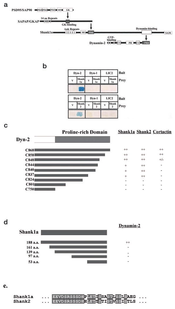
a, chain of interactions involving Shank1 and dynamin. Interaction domains identified in previous studies or in this paper are indicated by bold arrows. SH, SH3 domain; GK, guanylate kinase domain; Ank, ankyrin repeats; CC, coiled-coil domain; PRD, proline-rich domain. b, yeast two-hybrid assay for dynamin interactions. Bait constructs were full-length wild-type dynamin-2 (Dyn-2), dynamin-1 (Dyn-1), and cytoplasmic dynein light intermediate chain 2 (LIC2). Prey constructs were the initially isolated 188-a.a. Shank1 fragment, full-length Shank2, and the JG4–5 vector (V). c, mapping of the Shank1, Shank2, and cortactin binding sites within dynamin-2. Carboxyl-terminal deletions of the dynamin-2 proline-rich domain were used as bait in yeast two-hybrid assays with Shank1, Shank2, and cortactin as prey. ++, very strong interaction; +, strong interaction; +/–, weak but detectable interaction; −, undetectable interaction. d, mapping of the dynamin-2 binding sites in Shank1. Full-length and amino-terminal deletions of the original 188-a.a. Shank1 prey fragment were assayed for interaction with the dynamin-2 bait in a yeast two-hybrid assay. e, sequence comparison between Shank1 and Shank2 using the Clustal W sequence alignment method of the putative dynamin-2 binding site. Within this 29-a.a. region, 72% of the amino acids are identical as highlighted.
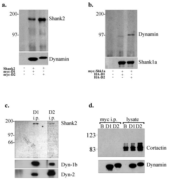
a, co-expression of full-length Shank2 with Myc-dynamin-1 or -2 in COS-7 cells. The dynamin isoforms were immunoprecipitated with anti-Myc, and the immunoprecipitates were immunoblotted with anti-Shank2 and anti-Myc. b, co-expression of Myc-tagged Shank1 with HA-tagged dynamin-1 or -2 in COS 7 cells. Shank1 was immunoprecipitated with anti-Myc, and the immunoprecipitates were blotted with anti-Myc and anti-HA antibodies. c, immunoprecipitation (i.p.) of dynamin-1 and -2 from rat brain cytosol using isoform-specific antibodies. The immunoprecipitates were blotted using anti-Shank2, anti-dynamin-1, and anti-dynamin-2 antibodies as indicated at right. d, anti-Myc immunoprecipitation of Myc-dynamin-1 and -2 singly expressed in COS-7 cells. The immunoprecipitates were detected using anti-Myc or anti-cortactin and showed no evidence for co-precipitation of cortactin with dynamin. B, beads alone control; D1, dynamin-1; D2, dynamin-2.
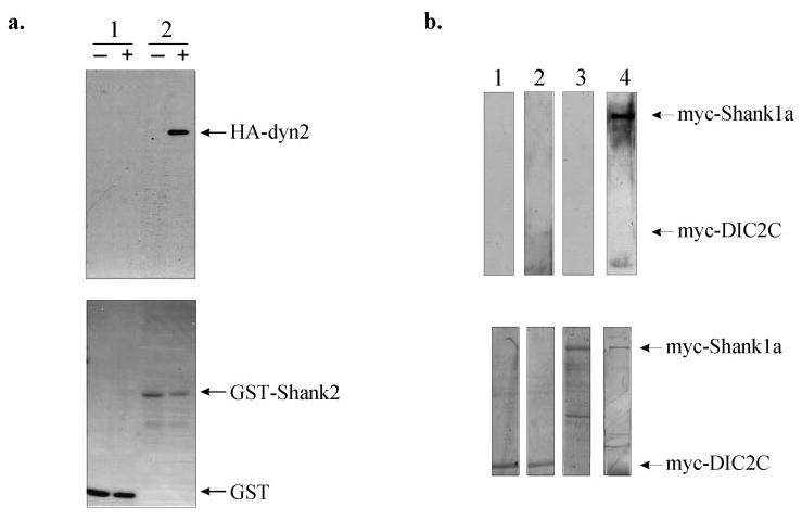
a, GST pull-down assays. Sepharose 4B beads, charged with either the GST-Shank2 fusion protein or GST alone, were incubated with purified recombinant HA-dynamin-2 and immunoblotted with an HA antibody. The upper panel shows that dynamin-2 directly interacts with GST-Shank2 (lane 2) but not with GST (lane 1), whereas the lower panel is the Coomassie-stained blot of the various purified GST proteins. −, absence of HA-dynamin-2; +, presence of HA-dynamin-2. b, blot overlay of Myc-tagged dynein intermediate chain 2C (DIC2C) or Myc-tagged Shank1 with purified recombinant HA-tagged dynamin-2. Shank1 and DIC2C were immunoprecipitated with an anti-Myc antibody and, after renaturation on the blot, overlaid with purified HA-dynamin-2 protein, which was detected with an anti-HA antibody. Upper panel, lanes 1 and 3, Myc-DIC2C and Myc-Shank1, respectively, in the absence of HA-dynamin-2; lanes 2 and 4, Myc-DIC2C and Myc-Shank1, respectively, overlaid with HA-dynamin-2. Lower panel, Coomassie-stained blots of the immunoprecipitated Myc-tagged proteins showing the location of each protein on the blot.
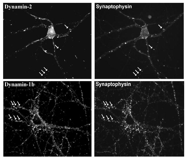
Rat hippocampal neurons (19 days in vitro) were double-labeled with anti-dynamin-1 or -2 antibodies versus anti-synaptophysin. Dynamin-2 exhibited punctate staining, which clearly overlapped with a subset of synaptophysin-positive spots (arrows). As a control, dynamin-1 also showed apparent co-localization with synaptophysin.
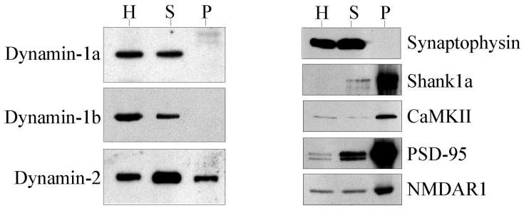
Adult rat brain homogenate (H), synaptosomal (S), and postsynaptic density (P) fractions were immunoblotted and probed with anti-dynamin-1a, -1b, and -2, anti-synaptophysin (a presynaptic marker), and anti-Shank1, anti-calmodulin kinase II, anti-PSD-95, and anti-NMDAR1 (PSD markers).
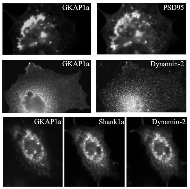
When dynamin-2 was co-expressed with two components of the postsynaptic density, GKAP1a and Shank1a/ProSAP, it co-localized into clusters that were mediated by its interaction with Shank1a/ProSAP.
References
Publication types
MeSH terms
Substances
Grants and funding
LinkOut - more resources
Full Text Sources
Molecular Biology Databases
