Review
. 2001 Oct;14(4):778-809, table of contents.
doi: 10.1128/CMR.14.4.778-809.2001.

Antiviral actions of interferons

Affiliations
Review

Antiviral actions of interferons

C E Samuel. Clin Microbiol Rev. 2001 Oct.

Abstract

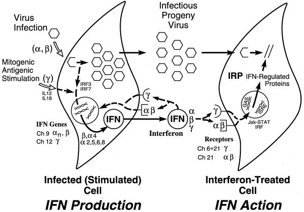
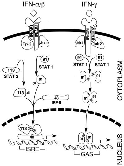
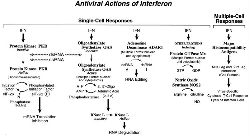
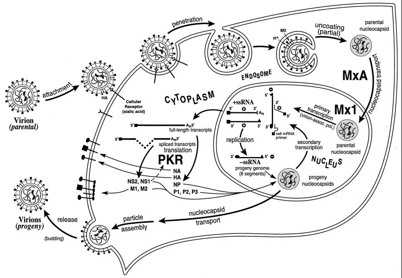
Tremendous progress has been made in understanding the molecular basis of the antiviral actions of interferons (IFNs), as well as strategies evolved by viruses to antagonize the actions of IFNs. Furthermore, advances made while elucidating the IFN system have contributed significantly to our understanding in multiple areas of virology and molecular cell biology, ranging from pathways of signal transduction to the biochemical mechanisms of transcriptional and translational control to the molecular basis of viral pathogenesis. IFNs are approved therapeutics and have moved from the basic research laboratory to the clinic. Among the IFN-induced proteins important in the antiviral actions of IFNs are the RNA-dependent protein kinase (PKR), the 2',5'-oligoadenylate synthetase (OAS) and RNase L, and the Mx protein GTPases. Double-stranded RNA plays a central role in modulating protein phosphorylation and RNA degradation catalyzed by the IFN-inducible PKR kinase and the 2'-5'-oligoadenylate-dependent RNase L, respectively, and also in RNA editing by the IFN-inducible RNA-specific adenosine deaminase (ADAR1). IFN also induces a form of inducible nitric oxide synthase (iNOS2) and the major histocompatibility complex class I and II proteins, all of which play important roles in immune response to infections. Several additional genes whose expression profiles are altered in response to IFN treatment and virus infection have been identified by microarray analyses. The availability of cDNA and genomic clones for many of the components of the IFN system, including IFN-alpha, IFN-beta, and IFN-gamma, their receptors, Jak and Stat and IRF signal transduction components, and proteins such as PKR, 2',5'-OAS, Mx, and ADAR, whose expression is regulated by IFNs, has permitted the generation of mutant proteins, cells that overexpress different forms of the proteins, and animals in which their expression has been disrupted by targeted gene disruption. The use of these IFN system reagents, both in cell culture and in whole animals, continues to provide important contributions to our understanding of the virus-host interaction and cellular antiviral response.


Figures

FIG. 1
FIG. 1
Schematic summary of the IFN system. Virion particles are illustrated as open hexagons, and IFN proteins are illustrated as open circles. The IFN-producing cell shown on the left depicts a cell induced to synthesize IFN in response to either virus infection (IFN-α and IFN-β) or antigen or mitogen stimulation (IFN-γ). The IFN-treated cell shown on the right depicts paracrine IFN action in a cell induced to synthesize IFN-regulated proteins that collectively constitute the antiviral response that is responsible for the inhibition of virus multiplication. IFN may also act in an autocrine manner on the IFN producer cell. The numbers refer to the chromosome (Ch) assignment of the human genes encoding the IFNs and their receptors. Adapted from reference with permission of the publisher.
FIG. 2
FIG. 2
Schematic of IFN signaling by the Jak-Stat pathway. The signaling process is initiated by binding of the IFN ligand to its cognate receptor subunits. IFN-α and IFN-β are depicted by the diamond, and IFN-γ is depicted by the sphere. IFN binding leads to activation of overlapping pairs of Jak and Stat transcription factors by tyrosine phosphorylation. The Jak-1 and Tyk-2 kinases are activated by IFN-α/β, which leads to the phosphorylation and dimerization of the Stat-1 (p91) and Stat-2 (p113) proteins and subsequent translocation, along with IRF-9 (p48), to the nucleus. The complex of these three proteins, known as IFN-stimulated gene factor 3 (ISGF-3), activates the transcription of IFN-α/β–inducible genes through the ISRE. The Jak-1 and Jak-2 kinases are activated by IFN-γ, which leads to the phosphorylation and homodimerization of the Stat-1 protein and subsequent translocation to the nucleus. The Stat-1 dimer complex, known as GAF for gamma activation factor, activates the transcription of IFN-γ inducible genes through the GAS enhancer element.
FIG. 3
FIG. 3
Functions of selected IFN-inducible proteins. Among the IFN-induced proteins believed to affect virus multiplication within single cells are PKR kinase, which inhibits translation initiation through the phosphorylation of protein synthesis initiation factor eIF-2α; the OAS synthetase family and RNase L nuclease, which mediate RNA degradation; the family of Mx protein GTPases, which appear to target viral nucleocapsids and inhibit RNA synthesis; and ADAR, which edits double-stranded RNA by deamination of adenosine to yield inosine. IFN-induced expression of MHC class I and class II antigens and NOS may contribute to the antiviral responses observed within whole animals. Ag, antigen. Adapted from reference with permission of the publisher.
FIG. 4
FIG. 4
Schematic diagram of the influenza virus multiplication cycle including sites of action of the IFN-induced Mx and PKR proteins. Enveloped influenza virus virion particles are depicted as spheres, in which the viral HA glycoprotein mediates binding to the cellular receptor. Virus penetration via receptor-mediated endocytosis is followed by partial uncoating and release of the viral nucleocapsid into the cytoplasm. The eight minus-strand RNA genome segments are shown as wavy lines in the nucleocapsid structures. The parental nucleocapsid is imported into the nucleus, where primary transcription is catalyzed by the virion-associated polymerase. Influenza virus transcripts are processed and transported to the cytoplasm, where they are translated by the host cell protein-synthesizing machinery. The IFN-induced PKR protein kinase that inhibits translation by phosphorylation of eIF-2, can be antagonized by the accumulated viral NS1 protein that binds dsRNA. The IFN-induced family of Mx antiviral proteins appear to act by affecting viral nucleocapsid transport and RNA synthesis. Human MxA accumulates in the cytoplasm, whereas mouse Mx1 accumulates in the nucleus of IFN-treated cells. The GTPase activity of Mx proteins is required for their antiviral activity. Newly synthesized progeny nucleocapsids may participate in further transcription or may be exported to the cytoplasm, where particle assembly occurs at the plasma membrane, with release of the enveloped progeny virions by a budding process.
FIG. 5
FIG. 5
Antagonism of the antiviral actions of IFN by viral gene products. Virus-encoded products that antagonize the IFN signaling pathway leading to transcriptional activation of IFN-regulated genes include adenovirus E1A; EBV EBNA2; HHV-8 K9; HPV E6; poxvirus B8R, B18R, and M-T7; SV5 V protein; and SeV C proteins. Virus-encoded RNA and protein products that antagonize the biochemical activity of IFN-induced cellular proteins include adenovirus VA RNA, EBV EBER RNA, HCV NS5a and E2 proteins, HSV γ34.5 and Us11 proteins, influenza virus NS1 protein, and SV40 T-antigen.
FIG. 6
FIG. 6
Schematic diagram of the vaccinia virus multiplication cycle including sites of action of poxvirus-encoded antagonists of the IFN response. The enveloped vaccinia virion particle has a large linear dsDNA genome. Virus penetration probably involves fusion between the viral envelope and cellular membranes, with release of the subviral core particle into the cytoplasm, where virus replication occurs. Early viral mRNAs synthesized by the virion-associated RNA polymerase include viral transcripts that encode IFN system antagonists. The E3L RNA-binding protein impairs activation of the PKR kinase and the OAS synthetases, IFN-induced enzymes that are activated by dsRNA. K3L is a substrate homologue of eIF-2α and is believed to antagonize the translation inhibition mediated by PKR. The B8R and B18R proteins are soluble IFN-γ and IFN-α/β receptor homologues, respectively, that serve as decoys to bind the extracellular induced IFNs. Early viral gene products include proteins and enzymes needed for further uncoating and for viral DNA replication and intermediate transcription. Late genes transcribed after DNA replication encode the structural proteins and enzymes necessary for progeny virion formation in the cytoplasm.

References

    1. Abendroth A, Slobedman B, Lee E, Mellins E, Wallace M, Arvin A M. Modulation of major histocompatibility class II protein expression by varicella-zoster virus. J Virol. 2000;74:1900–1907. - PMC - PubMed
    1. Abraham N, Stojdl D F, Duncan P I, Méthot N, Ishii T, Dubé M, Vanderhyden B C, Atkins H L, Gray D A, McBurney M W, Koromilas A E, Brown E G, Sonenberg N, Bell J C. Characterization of transgenic mice with targeted disruption of the catalytic domain of the double-stranded RNA-dependent protein kinase, PKR. J Biol Chem. 1999;274:5953–5962. - PubMed
    1. Aebi M, Fäh J, Hurt N, Samuel C E, Thomis D, Bazzigher L, Pavlovic J, Haller O, Staeheli P. cDNA Structures and regulation of two interferon-induced human Mx proteins. Mol Cell Biol. 1989;9:5062–5072. - PMC - PubMed
    1. Alberts B, Bray D, Lewis J, Raff M, Roberts K, Watson J D. Molecular biology of the cell. 3rd ed. New York, N.Y: Garland Publishing; 1994.
    1. Alcami A, Symons J, Smith G L. The vaccinia virus soluble alpha/beta interferon receptor binds to the cell surface and protects cells from the antiviral effects of IFN. J Virol. 2000;74:11230–11239. - PMC - PubMed

Publication types

MeSH terms