. 2001 Nov;21(21):7163-71.
doi: 10.1128/MCB.21.21.7163-7171.2001.

Stress-specific activation and repression of heat shock factors 1 and 2

Affiliations

Stress-specific activation and repression of heat shock factors 1 and 2

A Mathew et al. Mol Cell Biol. 2001 Nov.

Abstract

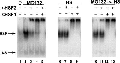
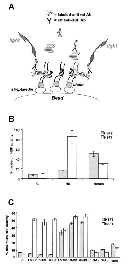
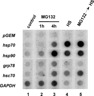
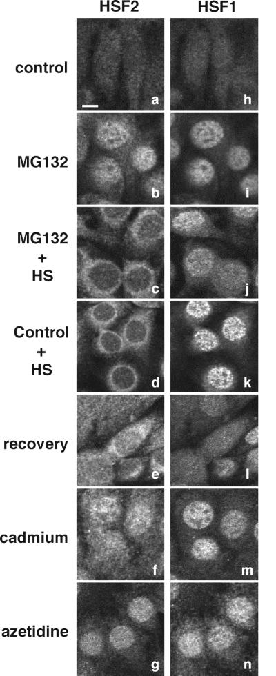
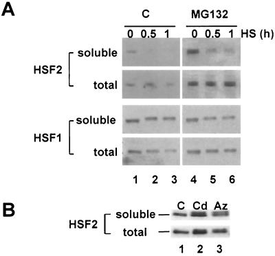
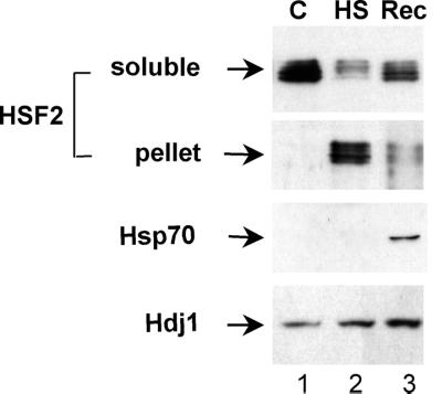
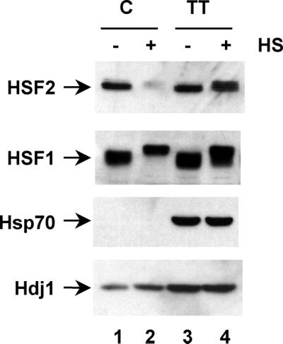
Vertebrate cells express a family of heat shock transcription factors (HSF1 to HSF4) that coordinate the inducible regulation of heat shock genes in response to diverse signals. HSF1 is potent and activated rapidly though transiently by heat shock, whereas HSF2 is a less active transcriptional regulator but can retain its DNA binding properties for extended periods. Consequently, the differential activation of HSF1 and HSF2 by various stresses may be critical for cells to survive repeated and diverse stress challenges and to provide a mechanism for more precise regulation of heat shock gene expression. Here we show, using a novel DNA binding and detection assay, that HSF1 and HSF2 are coactivated to different levels in response to a range of conditions that cause cell stress. Above a low basal activity of both HSFs, heat shock preferentially activates HSF1, whereas the amino acid analogue azetidine or the proteasome inhibitor MG132 coactivates both HSFs to different levels and hemin preferentially induces HSF2. Unexpectedly, we also found that heat shock has dramatic adverse effects on HSF2 that lead to its reversible inactivation coincident with relocalization from the nucleus. The reversible inactivation of HSF2 is specific to heat shock and does not occur with other stressors or in cells expressing high levels of heat shock proteins. These results reveal that HSF2 activity is negatively regulated by heat and suggest a role for heat shock proteins in the positive regulation of HSF2.


Figures

FIG. 1
FIG. 1
Effects of heat shock and MG132 treatments on activation of HSF1 and HSF2. Gel mobility shift analyses using extracts from untreated 3T3 control cells (C) (lane 1) or cells treated with MG132 (lanes 2 to 5), heat shock (HS) (lanes 6 to 9), or MG132 followed by heat shock (lanes 10 to 13) are shown. Cell extracts were incubated in either the presence or absence of specific antiserum to HSF1 or HSF2 or a mixture of both antisera, as indicated, prior to the gel mobility shift assay. HSF DNA binding activities and nonspecific binding (NS) are indicated by arrows. The radiolabeled complex detected at the top of the gel corresponds to retarded antibody-HSF complexes.
FIG. 2
FIG. 2
ORIGEN-based assay of HSF DNA binding activity. (A) Assay scheme as described in the text. Ab, antibody. (B) ORIGEN-based detection of HSF1 and HSF2 activities in K562 cells. Soluble whole-cell extracts (20 μg of protein) from control (C), heat-shock treated (HS), or hemin-induced K562 cells were incubated with 0.05 μg of rat anti-mouse HSF1 (4B4) or HSF2 (3E2) antibodies. The extracts were simultaneously incubated with biotinylated double-stranded HSE-containing oligonucleotide and an ORIGEN tag-labeled goat anti-rat antibody. The complex was allowed to bind to streptavidin-coated Dynal beads and used to measure the amount of bound HSF as described in the text. The sum of the HSF1 and HSF2 signals determined total HSF activity. The data are presented as percent maximum HSF activity, where maximum is defined by the condition which resulted in the highest total HSF activation (heat shock in this case). The data are derived from triplicate samples with coeffi- cients of variation of below 15%. Error bars indicate standard deviations. (C) HSF1 and HSF2 activities induced by heat, MG132, or azetidine treatment of 3T3 cells. 3T3 cells were treated for the lengths of time indicated with heat at 43°C (HS), 10 μM MG132 (MG), or 5 mM azetidine (Az). Extracts were prepared and used in the ORIGEN assay as described in the text. The data are derived from duplicate samples with coefficients of variation of 10% or less and are presented as percent maximum HSF activity as defined above, with maximum activity being induced in this case by the 6-h MG132 treatment. The data for both ORIGEN assays are corrected for nonspecific binding of tagged antibody determined by parallel assays containing all components except the primary antibodies.
FIG. 3
FIG. 3
Effects of heat shock and MG132 on rates of transcription of heat shock genes. Nuclear run-on analyses of hsp70, hsp90, grp78, hsc70, and GAPDH gene transcription in control 3T3 cells (lane 1) or cells treated with MG132 for 1 and 4 h (lanes 2 and 3, respectively), heat shock (HS) at 43°C for 0.5 h (lane 4), or MG132 for 4 h followed by heat shock at 43°C for 0.5 h (lane 5) are shown. The vector corresponds to the plasmid pGEM4, used to determine level of nonspecific hybridization. The levels of transcription were determined relative to nonspecific background and the internal control, the GAPDH gene.
FIG. 4
FIG. 4
Stress-dependent changes in intracellular localization of HSF2 and HSF1. Double immunofluorescence analyses were performed using specific antibodies to HSF2 (a to g) and HSF1 (h to n) to determine their intracellular localization in control 3T3 cells (a and h) or cells treated with MG132 (4 h) (b and i), MG132 (4 h) followed by heat shock (1.5 h) (c and j), heat shock (1.5 h) (d and k), heat shock (1.5 h) followed by 6 h of recovery at 37°C (e and l), cadmium (4 h) (f and m), or azetidine (4 h) (g and n). Bar, 5 μm.
FIG. 5
FIG. 5
Heat shock affects the solubility of HSF2. (A) Western blot analyses of HSF2 and HSF1 proteins in soluble and total cell extracts from control (lanes 1 to 3) or MG132-treated (lanes 4 to 6) 3T3 cells exposed to heat shock for 0 (lanes 1 and 4), 0.5 (lanes 2 and 5), or 1 (lanes 3 and 6) h. (B) Western blot analyses of HSF2 protein present in soluble or total extracts from control 3T3 cells (C) or cells treated with cadmium (Cd) or azetidine (Az) for 2 h.
FIG. 6
FIG. 6
HSF2 levels recover following heat shock. Western blot analysis of HSF2, Hsp70, and Hdj1 in the soluble fractions (first, third and fourth panels) and pellet fractions from control 3T3 cells (C) (lane 1) or cells exposed to heat shock (HS) (lane 2) and allowed to recover for 6 h at 37°C (Rec) (lane 3) is shown.
FIG. 7
FIG. 7
HSF2 is refractile to heat shock in thermotolerant cells. HSF2, HSF1, Hsp70, and Hdj1 were detected by Western blot analysis of soluble extracts from control (C) (lanes 1 and 2) or thermotolerant (TT) 3T3 cells treated with (lanes 2 and 4) or without (lanes 1 and 3) heat shock (HS).

References

    1. Ali A, Bharadwaj S, O'Carroll R, Ovsenek N. HSP90 interacts with and regulates the activity of heat shock factor 1 in Xenopus oocytes. Mol Cell Biol. 1998;18:4949–4960. - PMC - PubMed
    1. Baler R, Dahl G, Voellmy R. Activation of human heat shock genes is accompanied by oligomerization, modification, and rapid translocation of heat shock transcription factor HSF1. Mol Cell Biol. 1993;13:2486–2496. - PMC - PubMed
    1. Banerji S S, Theodorakis N G, Morimoto R I. Heat shock-induced translational control of HSP70 and globin synthesis in chicken reticulocytes. Mol Cell Biol. 1984;4:2437–2448. - PMC - PubMed
    1. Cotto J, Fox S S, Morimoto R. HSF1 granules: a novel stress-induced nuclear compartment of human cells. J Cell Sci. 1997;110:2925–2934. - PubMed
    1. Czarnecka-Verner E, Yuan C X, Fox P C, Gurley W B. Isolation and characterization of six heat shock transcription factor cDNA clones from soybean. Plant Mol Biol. 1995;29:37–51. - PubMed

Publication types

LinkOut - more resources