. 2001 Nov;21(21):7218-30.
doi: 10.1128/MCB.21.21.7218-7230.2001.

Transforming growth factor beta1 (TGF-beta1) promotes endothelial cell survival during in vitro angiogenesis via an autocrine mechanism implicating TGF-alpha signaling

Affiliations

Transforming growth factor beta1 (TGF-beta1) promotes endothelial cell survival during in vitro angiogenesis via an autocrine mechanism implicating TGF-alpha signaling

F Viñals et al. Mol Cell Biol. 2001 Nov.

Abstract

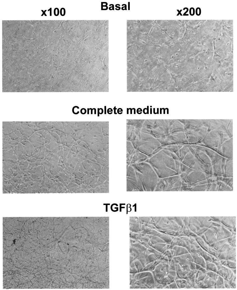
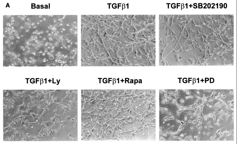
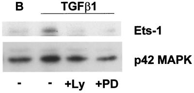
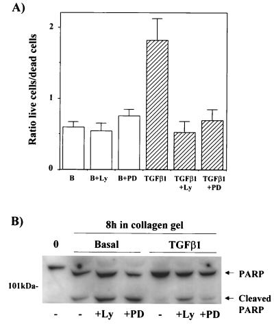
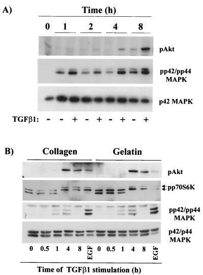
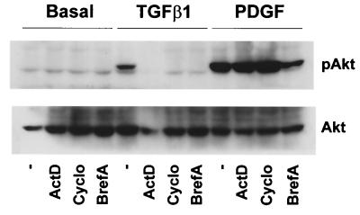
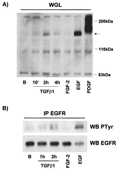
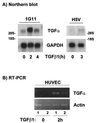
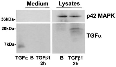
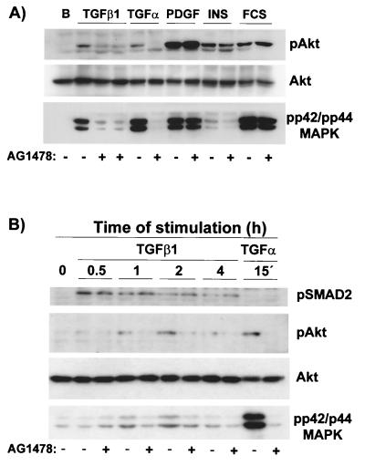
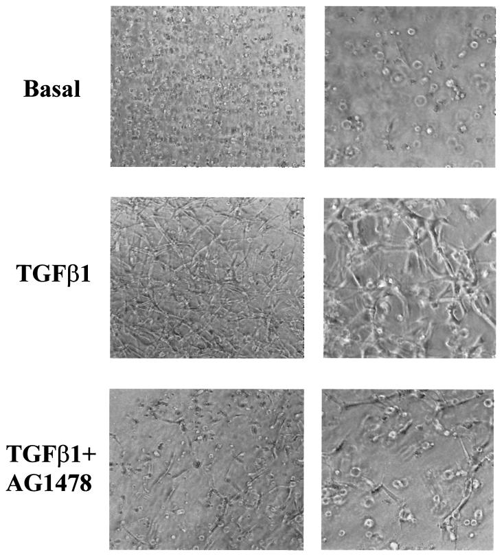
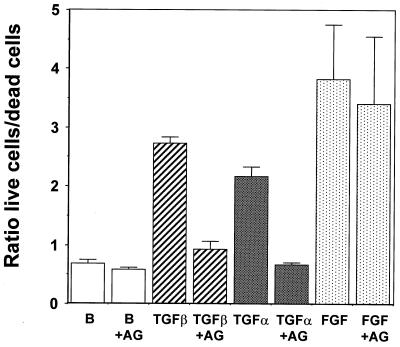
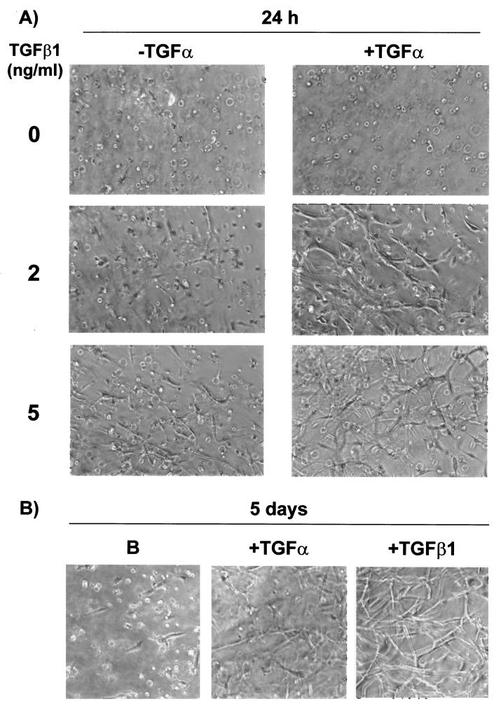
Mouse capillary endothelial cells (1G11 cell line) embedded in type I collagen gels undergo in vitro angiogenesis. Cells rapidly reorganize and form capillary-like structures when stimulated with serum. Transforming growth factor beta1 (TGF-beta1) alone can substitute for serum and induce cell survival and tubular network formation. This TGF-beta1-mediated angiogenic activity depends on phosphatidylinositol 3-kinase (PI3K) and p42/p44 mitogen-activated protein kinase (MAPK) signaling. We showed that specific inhibitors of either pathway (wortmannin, LY-294002, and PD-98059) all suppressed TGF-beta1-induced angiogenesis mainly by compromising cell survival. We established that TGF-beta1 stimulated the expression of TGF-alpha mRNA and protein, the tyrosine phosphorylation of a 170-kDa membrane protein representing the epidermal growth factor (EGF) receptor, and the delayed activation of PI3K/Akt and p42/p44 MAPK. Moreover, we showed that all these TGF-beta1-mediated signaling events, including tubular network formation, were suppressed by incubating TGF-beta1-stimulated endothelial cells with a soluble form of an EGF receptor (ErbB-1) or tyrphostin AG1478, a specific blocker of EGF receptor tyrosine kinase. Finally, addition of TGF-alpha alone poorly stimulated angiogenesis; however, by reducing cell death, it strongly potentiated the action of TGF-beta1. We therefore propose that TGF-beta1 promotes angiogenesis at least in part via the autocrine secretion of TGF-alpha, a cell survival growth factor, activating PI3K/Akt and p42/p44 MAPK.


Figures

FIG. 1
FIG. 1
TGF-β1 and complete medium stimulate 1G11 capillary endothelial cells to form a tubular network in type I collagen gels. 1G11 cells were mixed with type I collagen and placed on culture plates to polymerize. After gel formation, DMEM alone (basal), complete medium (20% FCS and 150 μg of endothelial cell growth supplement/ml), or TGF-β1 (10 ng/ml) was added for 48 h and gels were examined by phase-contrast microscopy.
FIG. 2
FIG. 2
LY-294002 and PD-98059 prevent TGF-β1-induced tube formation and Ets-1 induction. (A) 1G11 endothelial cells immersed in collagen gels were cultured for 24 h in the presence of DMEM alone (basal) or 10 ng of TGF-β1/ml either alone or with 10 μM SB202190, 15 μM LY-294002 (Ly), 10 nM rapamycin (rapa), or 30 μM PD-98059 (PD). Cells were examined by phase-contrast microscopy. Magnification, ×128. (B) 1G11 cells grown in collagen gels for 24 h in the presence of 10 ng of TGF-β1/ml either alone (TGF-β1 lane −) or with 15 μM LY-294002 (lane +Ly) or 30 μM PD-98059 (lane +PD) or in the absence of TGF-β (B lane −) were lysed, and Ets-1 was detected by immunoblotting with a specific antibody. Identical amounts of protein were loaded on the gel. The decrease in p42 MAPK content in lanes +Ly and +PD reflects partial cell death. A representative Western blot of three different experiments is shown.
FIG. 2
FIG. 2
LY-294002 and PD-98059 prevent TGF-β1-induced tube formation and Ets-1 induction. (A) 1G11 endothelial cells immersed in collagen gels were cultured for 24 h in the presence of DMEM alone (basal) or 10 ng of TGF-β1/ml either alone or with 10 μM SB202190, 15 μM LY-294002 (Ly), 10 nM rapamycin (rapa), or 30 μM PD-98059 (PD). Cells were examined by phase-contrast microscopy. Magnification, ×128. (B) 1G11 cells grown in collagen gels for 24 h in the presence of 10 ng of TGF-β1/ml either alone (TGF-β1 lane −) or with 15 μM LY-294002 (lane +Ly) or 30 μM PD-98059 (lane +PD) or in the absence of TGF-β (B lane −) were lysed, and Ets-1 was detected by immunoblotting with a specific antibody. Identical amounts of protein were loaded on the gel. The decrease in p42 MAPK content in lanes +Ly and +PD reflects partial cell death. A representative Western blot of three different experiments is shown.
FIG. 3
FIG. 3
TGF-β1 stimulates endothelial cell survival in collagen gels. (A) 1G11 cells immersed in collagen gels were cultured for 24 h in the presence of DMEM alone (B), 15 μM LY-294002 alone (B+Ly), 30 μM PD-98059 alone (B+PD), 10 ng of TGF-β1/ml (TGFβ1), and TGF-β1 in the presence of 15 μM LY-294002 (TGFβ1+Ly) or 30 μM PD-98059 (TGFβ1+PD). After this time, live and dead cells were counted as described in Materials and Methods. Results are expressed as the ratios of live cells/dead cells and are averages of eight different experiments. (B) Proliferative 1G11 cells (0) or cells immersed in collagen gels for 8 h in the absence or presence of TGF-β1 (10 ng/ml), LY-294002 (15 μM), or PD-98059 (30 μM) were lysed, and PARP was detected by immunoblotting with a specific antibody. A representative Western blot is shown. The observed difference in the mobility of PARP between proliferative cells and cells immersed in collagen gels is due to the presence of collagen in the SDS-polyacrylamide gel electrophoresis.
FIG. 4
FIG. 4
TGF-β1 stimulates PI3K, p70 S6K, and p42/p44 MAPK activities in endothelial cells grown in collagen gels and in two-dimensional cultures. (A) 1G11 cells were grown in collagen gels for the periods of time indicated. Cells were lysed, and phospho-Akt, phospho-p42/p44 MAPK (pp42/pp44 MAPK), and p42 MAPK were detected by immunoblotting with specific antibodies. A representative Western blot is shown. (B) 1G11 cells were grown on collagen- or gelatin-coated plates until confluence. After depletion of growth factors, cells were stimulated with 10 ng of TGF-β1/ml for the periods of time indicated or with 20 ng of EGF for 30 min. Cells were lysed, and Western blotting was performed using anti-phospho-Akt, anti-phospho-p42/p44 MAPK, or p42/p44 MAPK. The same extracts were loaded on an SDS–9% polyacrylamide gel (shift-up) and blotted with an anti-p70 S6K antibody. Hyperphosphorylated and active forms of p70 S6K (arrows) migrated more slowly than hypophosphorylated forms. The Western blots are representative of three independent experiments.
FIG. 5
FIG. 5
Effect of TGF-β1 on Akt and p42/p44 MAPK activation depends on mRNA and protein synthesis and vesicular secretion. Quiescent 1G11 cells were preincubated for 15 min in the presence of 5 μg of actinomycin D/ml, 10 μg of cycloheximide/ml, or 1 μg of brefeldin A/ml or in the absence of inhibitors (−), followed by a 4-h stimulation with 10 ng of TGF-β1/ml, a 30-min stimulation with 10 ng of PDGF-BB/ml, or no stimulation (basal). Cells were lysed, and phospho-Akt and Akt were immunodetected as previously (76) described. A Western blot representative of three different experiments is shown.
FIG. 6
FIG. 6
Stimulation with TGF-β1 causes EGF receptor activation. (A) Quiescent 1G11 cells were stimulated or not (lane B) for the indicated times with 10 ng of TGF-β1/ml and for 10 min with 25 ng of FGF-2/ml, 10 ng of EGF/ml, or 10 ng of PDGF-BB/ml. Cells were lysed, and proteins were incubated with Sepharose-WGL for 1 h. After being washed, the final pellet was resuspended in Laemmli sample buffer and loaded on a SDS–7.5% polyacrylamide gel. Phosphotyrosine-containing proteins were immunodetected by using a specific antibody. Arrow, phosphotyrosine-containing protein that appeared after TGF-β1 treatment. A representative Western blot is shown. (B) Cells were treated as for panel A and lysed, and the EGF receptor (EGFR) was immunoprecipitated (IP) by incubation with a specific anti-EGF receptor antibody preadsorbed to protein A-Sepharose beads. After being washed the pellet was treated as for panel A. WB, Western blot.
FIG. 7
FIG. 7
TGF-β1 induces TGF-α mRNA expression in endothelial cells. (A) 1G11 and H5V cells stimulated with 10 ng of TGF-β1/ml for the indicated times or not stimulated were lysed, and poly(A)+ mRNA was isolated. Gels were loaded with 2 μg of mRNA, and Northern blotting was performed using a 190-bp PCR-amplified fragment as a TGF-α-specific probe. Rat GAPDH (glyceraldehyde-3-phosphate dehydrogenase) was used as a control. A representative result is shown. (B) HUVEC stimulated with 10 ng of TGF-β1/ml for 2 h or not stimulated were lysed, and poly(A)+ mRNA was isolated. After cDNA was obtained, PCR was performed using specific primers for human TGF-α or actin (as a control). Samples were loaded on a 2% agarose gel. 1 and 2, two independent preparations of HUVEC poly(A)+ for unstimulated and stimulated cells.
FIG. 8
FIG. 8
TGF-β1 induces TGF-α protein expression in 1G11 cells. Quiescent 1G11 cells were stimulated for 2 h with 10 ng of TGF-β1/ml or were not stimulated (lane B). After this time, medium was collected and proteins were precipitated and loaded on a 15% gel. As a control, 300 ng of human TGF-α was also loaded. In parallel, cells were lysed and 100 μg was loaded on the gel. TGF-α was immunodetected by using a specific antibody. A Western blot representative of three different experiments is shown.
FIG. 9
FIG. 9
Tyrphostin AG1478, a specific inhibitor of the EGF receptor, blocks TGF-β1 stimulation of p42/p44 MAPK and Akt in the absence of changes in TGF-β signaling. (A) Quiescent 1G11 cells were preincubated for 15 min in the presence (+) or the absence (−) of tyrphostin AG1478 (1 μM). After this time, cells were stimulated for 4 h with 10 ng of TGF-β1/ml (in duplicate in the presence of tyrphostin AG1478) or for 1 h with 50 ng of TGF-α/ml, 10 ng of PDGF-BB/ml, 1 μM insulin, or 10% FCS or were left unstimulated (lane B). Cells were lysed, and phospho-Akt, Akt, and phospho-p42/p44 MAPK were immunodetected as described in Materials and Methods. A Western blot representative of four different experiments is shown. (B) Quiescent 1G11 cells were preincubated for 15 min in the presence (+) or the absence (−) of tyrphostin AG1478 (1 μM). After this time, cells were stimulated for the times indicated with 10 ng of TGF-β1/ml or for 15 min with 50 ng of TGF-α/ml. After lysis, phospho-Smad2, phospho-Akt, Akt, and phospho-p42/p44 MAPK were immunodetected as described in Materials and Methods. A representative Western blot is shown.
FIG. 10
FIG. 10
A soluble EGF receptor (IgB-1) blocks TGF-β1 stimulation of Akt and p42/p44 MAPK. Quiescent 1G11 cells were preincubated for 1 h with medium from control or soluble extracellular EGF receptor IgB-1-transfected HEK293 cells. After this period, cells were stimulated with TGF-β1 for 4 h. Cells were then lysed, and phospho-Akt, phospho p42/p44 MAPK, and p42/p44 MAPK were immunodetected as described in Materials and Methods. A representative Western blot from three different experiments is shown.
FIG. 11
FIG. 11
Inhibition of TGF-α signaling blocks tube formation and cell survival induced by TGF-β1 in collagen gels. (Top) 1G11 endothelial cells grown in collagen gels were cultured for 24 h in the presence of DMEM alone (basal) or 10 ng of TGF-β1/ml either alone or supplemented with 1 μM tyrphostin AG1478. After this time, cells were examined by phase-contrast microscopy. Magnifications: left, ×90; right, ×180. (Bottom) 1G11 endothelial cells grown in collagen gels were cultured for 24 h in the presence of DMEM either alone (lane B) or with 1 μM tyrphostin AG1478 (B+AG), in 10 ng of TGF-β1/ml either alone (TGFβ) or with 1 μM tyrphostin AG1478 (TGFβ+AG), in 50 ng of TGF-α/ml either alone (TGFα) or with tyrphostin AG1478 (TGFα+AG), or in 100 ng of FGF-2/ml either alone (FGF) or with 1 μM tyrphostin AG1478 (FGF+AG). Dead and living cells were counted as described in Materials and Methods. Results are averages of three different experiments.
FIG. 11
FIG. 11
Inhibition of TGF-α signaling blocks tube formation and cell survival induced by TGF-β1 in collagen gels. (Top) 1G11 endothelial cells grown in collagen gels were cultured for 24 h in the presence of DMEM alone (basal) or 10 ng of TGF-β1/ml either alone or supplemented with 1 μM tyrphostin AG1478. After this time, cells were examined by phase-contrast microscopy. Magnifications: left, ×90; right, ×180. (Bottom) 1G11 endothelial cells grown in collagen gels were cultured for 24 h in the presence of DMEM either alone (lane B) or with 1 μM tyrphostin AG1478 (B+AG), in 10 ng of TGF-β1/ml either alone (TGFβ) or with 1 μM tyrphostin AG1478 (TGFβ+AG), in 50 ng of TGF-α/ml either alone (TGFα) or with tyrphostin AG1478 (TGFα+AG), or in 100 ng of FGF-2/ml either alone (FGF) or with 1 μM tyrphostin AG1478 (FGF+AG). Dead and living cells were counted as described in Materials and Methods. Results are averages of three different experiments.
FIG. 12
FIG. 12
TGF-α stimulates tube formation in collagen gels in the long term and potentiates TGF-β1 action in the short term. 1G11 cells grown in collagen gels were cultured for 24 h in the presence of DMEM alone or different concentrations of TGF-β1 (2 and 5 ng/ml) in the presence or the absence of TGF-α (50 ng/ml). In parallel, 1G11 cells were grown in collagen gels for 5 days in the presence or the absence of TGF-β1 (10 ng/ml) or TGF-α (50 ng/ml). Cells were examined by phase-contrast microscopy.

Similar articles

Cited by

References

    1. Alroy I, Yarden Y. The ErbB signaling network in embryogenesis and oncogenesis: signal diversification through combinatorial ligand-receptor interactions. FEBS Lett. 1997;410:83–86. - PubMed
    1. Arbiser J L, Moses M A, Fernandez C A, Ghiso N, Cao Y, Klauber N, Frank D, Brownlee M, Flynn E, Parangi S, Byers H R, Folkman J. Oncogenic H-ras stimulates tumor angiogenesis by two distinct pathways. Proc Natl Acad Sci USA. 1997;94:861–866. - PMC - PubMed
    1. Baird A, Durkin T. Inhibition of endothelial cell proliferation by type beta-transforming growth factor: interactions with acidic and basic fibroblast growth factors. Biochem Biophys Res Commun. 1986;138:476–482. - PubMed
    1. Banerjee S, Banerjee P P, Zirkin B R, Brown T R. Regional expression of transforming growth factor-alpha in rat ventral prostate during postnatal development, after androgen ablation, and after androgen replacement. Endocrinology. 1998;139:3005–3013. - PubMed
    1. Barbieri B, Balconi G, Dejana E, Donati M B. Evidence that vascular endothelial cells can induce the retraction of fibrin clots. Proc Soc Exp Biol Med. 1981;168:204–207. - PubMed

Publication types

MeSH terms

Substances